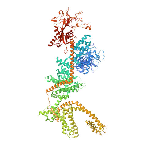
8SR7

Cryo-EM structure of TRPM2 chanzyme in the presence of Magnesium, Adenosine monophosphate, and Ribose-5-phosphate

  • Classification: TRANSPORT PROTEIN
  • Organism(s): Salpingoeca rosetta
  • Expression System: Homo sapiens
  • Mutation(s): No 
  • Membrane Protein: Yes  PDBTM

  • Deposited: 2023-05-05 Released: 2024-05-08 
  • Deposition Author(s): Huang, Y., Kumar, S., Lu, W., Du, J.
  • Funding Organization(s): National Institutes of Health/National Heart, Lung, and Blood Institute (NIH/NHLBI), National Institutes of Health/National Institute of Neurological Disorders and Stroke (NIH/NINDS), National Institutes of Health/National Institute of General Medical Sciences (NIH/NIGMS)

Experimental Data Snapshot

  • Method: ELECTRON MICROSCOPY
  • Resolution: 1.97 Å
  • Aggregation State: PARTICLE 
  • Reconstruction Method: SINGLE PARTICLE 

wwPDB Validation   3D Report Full Report


This is version 1.3 of the entry. See complete history


Literature

Coupling enzymatic activity and gating in an ancient TRPM chanzyme and its molecular evolution.

Huang, Y.Kumar, S.Lee, J.Lu, W.Du, J.

(2024) Nat Struct Mol Biol 31: 1509-1521

  • DOI: https://doi.org/10.1038/s41594-024-01316-4
  • Primary Citation of Related Structures:  
    8SR7, 8SR8, 8SR9, 8SRA, 8SRB, 8SRC, 8SRD, 8SRE, 8SRF, 8SRG, 8SRH, 8SRI, 8SRJ, 8SRK

  • PubMed Abstract: 

    Channel enzymes represent a class of ion channels with enzymatic activity directly or indirectly linked to their channel function. We investigated a TRPM2 chanzyme from choanoflagellates that integrates two seemingly incompatible functions into a single peptide: a channel module activated by ADP-ribose with high open probability and an enzyme module (NUDT9-H domain) consuming ADP-ribose at a remarkably slow rate. Using time-resolved cryogenic-electron microscopy, we captured a complete series of structural snapshots of gating and catalytic cycles, revealing the coupling mechanism between channel gating and enzymatic activity. The slow kinetics of the NUDT9-H enzyme module confers a self-regulatory mechanism: ADPR binding triggers NUDT9-H tetramerization, promoting channel opening, while subsequent hydrolysis reduces local ADPR, inducing channel closure. We further demonstrated how the NUDT9-H domain has evolved from a structurally semi-independent ADP-ribose hydrolase module in early species to a fully integrated component of a gating ring essential for channel activation in advanced species.


  • Organizational Affiliation

    Van Andel Institute, Grand Rapids, MI, USA.


Macromolecules
Find similar proteins by:  (by identity cutoff)  |  3D Structure
Entity ID: 1
MoleculeChains Sequence LengthOrganismDetailsImage
TRPM2 Chanzyme
A, B, C, D
1,494Salpingoeca rosettaMutation(s): 0 
Gene Names: PTSG_05449
Membrane Entity: Yes 
UniProt
Find proteins for F2UB89 (Salpingoeca rosetta (strain ATCC 50818 / BSB-021))
Explore F2UB89 
Go to UniProtKB:  F2UB89
Entity Groups  
Sequence Clusters30% Identity50% Identity70% Identity90% Identity95% Identity100% Identity
UniProt GroupF2UB89
Sequence Annotations
Expand
  • Reference Sequence
Small Molecules
Ligands 4 Unique
IDChains Name / Formula / InChI Key2D Diagram3D Interactions
CLR
Query on CLR

Download Ideal Coordinates CCD File 
DA [auth C]
EA [auth C]
FA [auth C]
GA [auth D]
HA [auth D]
DA [auth C],
EA [auth C],
FA [auth C],
GA [auth D],
HA [auth D],
K [auth A],
L [auth A],
M [auth A],
OA [auth D],
U [auth B],
V [auth B],
W [auth B]
CHOLESTEROL
C27 H46 O
HVYWMOMLDIMFJA-DPAQBDIFSA-N
AMP (Subject of Investigation/LOI)
Query on AMP

Download Ideal Coordinates CCD File 
F [auth A],
JA [auth D],
P [auth B],
Y [auth C]
ADENOSINE MONOPHOSPHATE
C10 H14 N5 O7 P
UDMBCSSLTHHNCD-KQYNXXCUSA-N
RP5 (Subject of Investigation/LOI)
Query on RP5

Download Ideal Coordinates CCD File 
E [auth A],
IA [auth D],
O [auth B],
X [auth C]
5-O-phosphono-beta-D-ribofuranose
C5 H11 O8 P
KTVPXOYAKDPRHY-TXICZTDVSA-N
MG (Subject of Investigation/LOI)
Query on MG

Download Ideal Coordinates CCD File 
AA [auth C]
BA [auth C]
CA [auth C]
G [auth A]
H [auth A]
AA [auth C],
BA [auth C],
CA [auth C],
G [auth A],
H [auth A],
I [auth A],
J [auth A],
KA [auth D],
LA [auth D],
MA [auth D],
N [auth A],
NA [auth D],
Q [auth B],
R [auth B],
S [auth B],
T [auth B],
Z [auth C]
MAGNESIUM ION
Mg
JLVVSXFLKOJNIY-UHFFFAOYSA-N
Experimental Data & Validation

Experimental Data

  • Method: ELECTRON MICROSCOPY
  • Resolution: 1.97 Å
  • Aggregation State: PARTICLE 
  • Reconstruction Method: SINGLE PARTICLE 
EM Software:
TaskSoftware PackageVersion
MODEL REFINEMENTPHENIX1.20.1_4487:

Structure Validation

View Full Validation Report


Entry History & Funding Information

Deposition Data


Funding OrganizationLocationGrant Number
National Institutes of Health/National Heart, Lung, and Blood Institute (NIH/NHLBI)United StatesHL153219
National Institutes of Health/National Institute of Neurological Disorders and Stroke (NIH/NINDS)United StatesNS112363
National Institutes of Health/National Institute of Neurological Disorders and Stroke (NIH/NINDS)United StatesNS111031
National Institutes of Health/National Institute of General Medical Sciences (NIH/NIGMS)United StatesGM129547

Revision History  (Full details and data files)

  • Version 1.0: 2024-05-08
    Type: Initial release
  • Version 1.1: 2024-06-05
    Changes: Database references
  • Version 1.2: 2024-10-23
    Changes: Data collection, Structure summary
  • Version 1.3: 2024-10-30
    Changes: Data collection, Database references