8SZK | pdb_00008szk

The cryo-EM structure of PPP2R5A/HIV-1 Vif/CBFb/EloB/EloC complex


Experimental Data Snapshot

  • Method: ELECTRON MICROSCOPY
  • Resolution: 3.58 Å
  • Aggregation State: PARTICLE 
  • Reconstruction Method: SINGLE PARTICLE 

wwPDB Validation 3D Report Full Report

Validation slider image for 8SZK

This is version 1.2 of the entry. See complete history

Literature

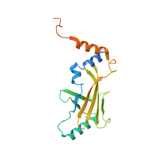
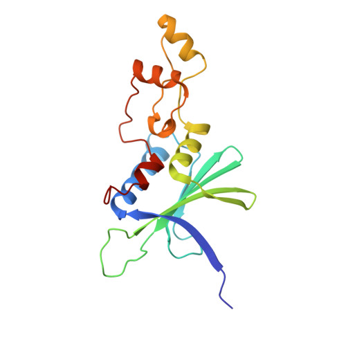
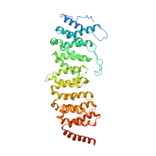
Structural insights into PPP2R5A degradation by HIV-1 Vif.

Hu, Y.Delviks-Frankenberry, K.A.Wu, C.Arizaga, F.Pathak, V.K.Xiong, Y.

(2024) Nat Struct Mol Biol 31: 1492-1501

  • DOI: https://doi.org/10.1038/s41594-024-01314-6
  • Primary Citation Related Structures: 
    8SZK

  • PubMed Abstract: 

    HIV-1 Vif recruits host cullin-RING-E3 ubiquitin ligase and CBFβ to degrade the cellular APOBEC3 antiviral proteins through diverse interactions. Recent evidence has shown that Vif also degrades the regulatory subunits PPP2R5(A-E) of cellular protein phosphatase 2A to induce G2/M cell cycle arrest. As PPP2R5 proteins bear no functional or structural resemblance to A3s, it is unclear how Vif can recognize different sets of proteins. Here we report the cryogenic-electron microscopy structure of PPP2R5A in complex with HIV-1 Vif-CBFβ-elongin B-elongin C at 3.58 Å resolution. The structure shows PPP2R5A binds across the Vif molecule, with biochemical and cellular studies confirming a distinct Vif-PPP2R5A interface that partially overlaps with those for A3s. Vif also blocks a canonical PPP2R5A substrate-binding site, indicating that it suppresses the phosphatase activities through both degradation-dependent and degradation-independent mechanisms. Our work identifies critical Vif motifs regulating the recognition of diverse A3 and PPP2R5A substrates, whereby disruption of these host-virus protein interactions could serve as potential targets for HIV-1 therapeutics.


  • Organizational Affiliation
    • Department of Molecular Biophysics and Biochemistry, Yale University, New Haven, CT, USA.

Macromolecule Content 

  • Total Structure Weight: 124.88 kDa 
  • Atom Count: 7,392 
  • Modeled Residue Count: 904 
  • Deposited Residue Count: 1,077 
  • Unique protein chains: 5

Macromolecules

Find similar proteins by:|  3D Structure
Entity ID: 1
MoleculeChains  Sequence LengthOrganismDetailsImage
Elongin-B118Homo sapiensMutation(s): 0 
Gene Names: ELOBTCEB2
UniProt & NIH Common Fund Data Resources
Find proteins for Q15370 (Homo sapiens)
Explore Q15370 
Go to UniProtKB:  Q15370
PHAROS:  Q15370
GTEx:  ENSG00000103363 
Entity Groups
Sequence Clusters30% Identity50% Identity70% Identity90% Identity95% Identity100% Identity
UniProt GroupQ15370
Sequence Annotations
Expand
Reference Sequence
Find similar proteins by:|  3D Structure
Entity ID: 2
MoleculeChains  Sequence LengthOrganismDetailsImage
Elongin-C96Homo sapiensMutation(s): 0 
Gene Names: ELOCTCEB1
UniProt & NIH Common Fund Data Resources
Find proteins for Q15369 (Homo sapiens)
Explore Q15369 
Go to UniProtKB:  Q15369
PHAROS:  Q15369
GTEx:  ENSG00000154582 
Entity Groups
Sequence Clusters30% Identity50% Identity70% Identity90% Identity95% Identity100% Identity
UniProt GroupQ15369
Sequence Annotations
Expand
Reference Sequence
Find similar proteins by:|  3D Structure
Entity ID: 3
MoleculeChains  Sequence LengthOrganismDetailsImage
Core-binding factor subunit beta201Homo sapiensMutation(s): 0 
Gene Names: CBFB
UniProt & NIH Common Fund Data Resources
Find proteins for Q13951 (Homo sapiens)
Explore Q13951 
Go to UniProtKB:  Q13951
PHAROS:  Q13951
GTEx:  ENSG00000067955 
Entity Groups
Sequence Clusters30% Identity50% Identity70% Identity90% Identity95% Identity100% Identity
UniProt GroupQ13951
Sequence Annotations
Expand
Reference Sequence
Find similar proteins by:|  3D Structure
Entity ID: 4
MoleculeChains  Sequence LengthOrganismDetailsImage
Virion infectivity factorD [auth G]176Human immunodeficiency virus type 1 (NEW YORK-5 ISOLATE)Mutation(s): 0 
Gene Names: vif
UniProt
Find proteins for P12504 (Human immunodeficiency virus type 1 group M subtype B (isolate NY5))
Explore P12504 
Go to UniProtKB:  P12504
Entity Groups
Sequence Clusters30% Identity50% Identity70% Identity90% Identity95% Identity100% Identity
UniProt GroupP12504
Sequence Annotations
Expand
Reference Sequence
Find similar proteins by:|  3D Structure
Entity ID: 5
MoleculeChains  Sequence LengthOrganismDetailsImage
Serine/threonine-protein phosphatase 2A 56 kDa regulatory subunit alpha isoformE [auth D]486Homo sapiensMutation(s): 0 
Gene Names: PPP2R5A
UniProt & NIH Common Fund Data Resources
Find proteins for Q15172 (Homo sapiens)
Explore Q15172 
Go to UniProtKB:  Q15172
PHAROS:  Q15172
GTEx:  ENSG00000066027 
Entity Groups
Sequence Clusters30% Identity50% Identity70% Identity90% Identity95% Identity100% Identity
UniProt GroupQ15172
Sequence Annotations
Expand
Reference Sequence

Experimental Data & Validation

Experimental Data

  • Method: ELECTRON MICROSCOPY
  • Resolution: 3.58 Å
  • Aggregation State: PARTICLE 
  • Reconstruction Method: SINGLE PARTICLE 
EM Software:
TaskSoftware PackageVersion
MODEL REFINEMENTPHENIX

Structure Validation

View Full Validation Report


Entry History 

& Funding Information

Deposition Data


Funding OrganizationLocationGrant Number
National Institutes of Health/National Cancer Institute (NIH/NCI)United States--

Revision History  (Full details and data files)

  • Version 1.0: 2024-06-05
    Type: Initial release
  • Version 1.1: 2024-10-30
    Changes: Data collection, Database references, Structure summary
  • Version 1.2: 2025-05-28
    Changes: Data collection