8T7H | pdb_00008t7h

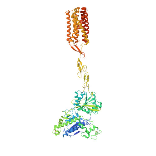
Quis-bound intermediate mGlu5


Experimental Data Snapshot

  • Method: ELECTRON MICROSCOPY
  • Resolution: 3.30 Å
  • Aggregation State: PARTICLE 
  • Reconstruction Method: SINGLE PARTICLE 

wwPDB Validation 3D Report Full Report

Validation slider image for 8T7H

This is version 1.4 of the entry. See complete history. 

Literature

Stepwise activation of a metabotropic glutamate receptor.

Krishna Kumar, K., Wang, H., Habrian, C., Latorraca, N.R., Xu, J., O'Brien, E.S., Zhang, C., Montabana, E., Koehl, A., Marqusee, S., Isacoff, E.Y., Kobilka, B.K.

(2024) Nature 629: 951-956

  • DOI: https://doi.org/10.1038/s41586-024-07327-x
  • Primary Citation Related Structures: 
    8T6J, 8T7H, 8T8M, 8TAO

  • PubMed Abstract: 

    Metabotropic glutamate receptors belong to a family of G protein-coupled receptors that are obligate dimers and possess a large extracellular ligand-binding domain that is linked via a cysteine-rich domain to their 7-transmembrane domain 1 . Upon activation, these receptors undergo a large conformational change to transmit the ligand binding signal from the extracellular ligand-binding domain to the G protein-coupling 7-transmembrane domain 2 . In this manuscript, we propose a model for a sequential, multistep activation mechanism of metabotropic glutamate receptor subtype 5. We present a series of structures in lipid nanodiscs, from inactive to fully active, including agonist-bound intermediate states. Further, using bulk and single-molecule fluorescence imaging, we reveal distinct receptor conformations upon allosteric modulator and G protein binding.


  • Organizational Affiliation: 
    • Department of Molecular and Cellular Physiology, Stanford University School of Medicine, Stanford, CA, USA. kaavyak@stanford.edu.

Macromolecule Content 

  • Total Structure Weight: 224.82 kDa 
  • Atom Count: 13,100 
  • Modeled Residue Count: 1,794 
  • Deposited Residue Count: 2,008 
  • Unique protein chains: 2

Macromolecules

Find similar proteins by:|  3D Structure
Entity ID: 1
MoleculeChains  Sequence LengthOrganismDetailsImage
Metabotropic glutamate receptor 5
A, B
881Homo sapiensMutation(s): 0 
Gene Names: GRM5, GPRC1E, MGLUR5
Membrane Entity: Yes 
UniProt & NIH Common Fund Data Resources
Find proteins for P41594 (Homo sapiens)
Explore P41594 
Go to UniProtKB:  P41594
PHAROS:  P41594
GTEx:  ENSG00000168959 
Entity Groups
Sequence Clusters30% Identity50% Identity70% Identity90% Identity95% Identity100% Identity
UniProt GroupP41594
Sequence Annotations
Expand
Reference Sequence
Find similar proteins by:|  3D Structure
Entity ID: 2
MoleculeChains  Sequence LengthOrganismDetailsImage
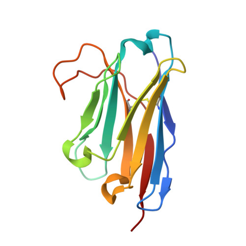
Nanobody 43
C, D
123Lama glamaMutation(s): 0 
Entity Groups
Sequence Clusters30% Identity50% Identity70% Identity90% Identity95% Identity100% Identity
Sequence Annotations
Expand
Reference Sequence

Small Molecules

Binding Affinity Annotations 
IDSourceBinding Affinity
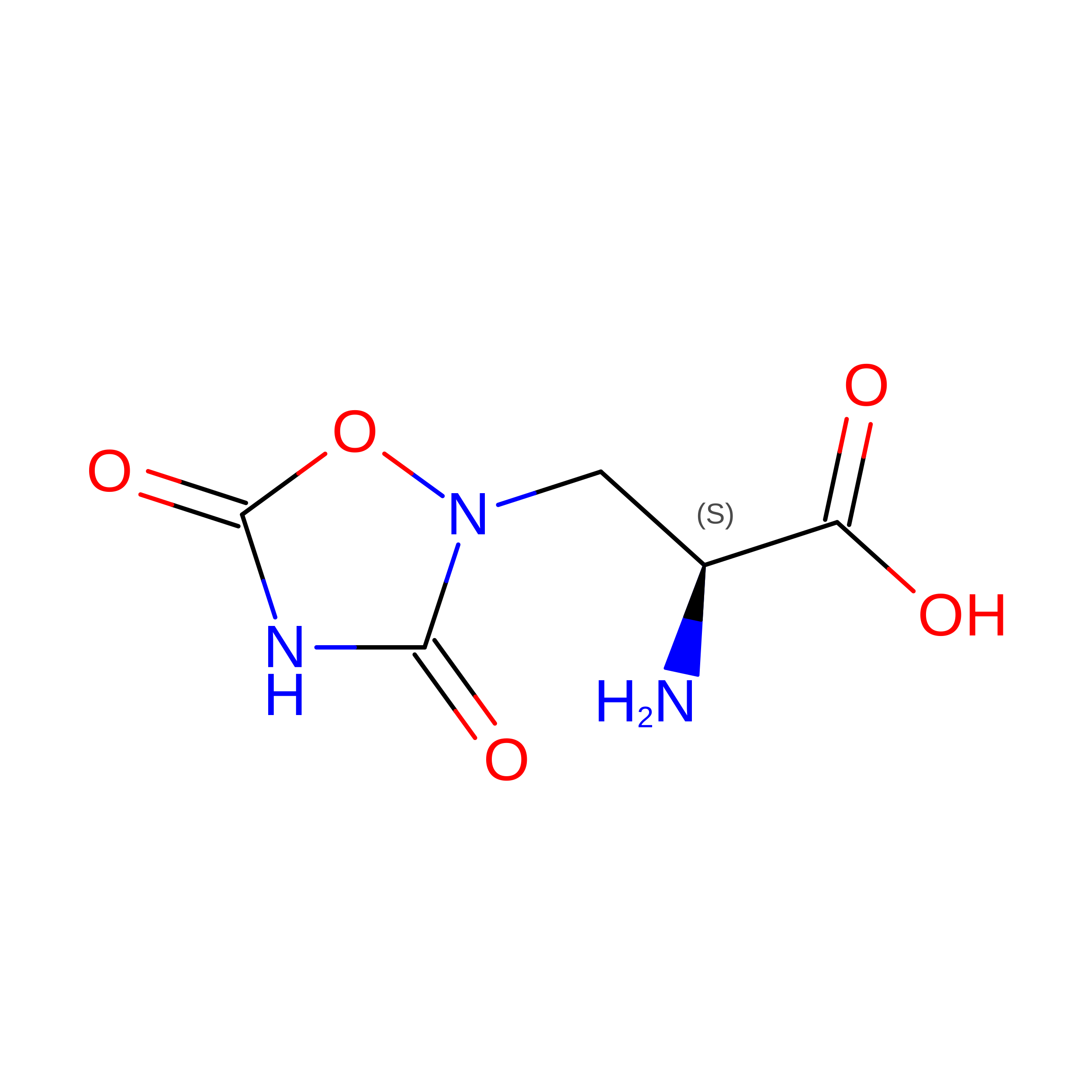
QUS BindingDB:  8T7H Ki: min: 1.00e+4, max: 1.00e+4 (nM) from 2 assay(s)

Experimental Data & Validation

Experimental Data

  • Method: ELECTRON MICROSCOPY
  • Resolution: 3.30 Å
  • Aggregation State: PARTICLE 
  • Reconstruction Method: SINGLE PARTICLE 
EM Software:
TaskSoftware PackageVersion
MODEL REFINEMENTPHENIX

Structure Validation

View Full Validation Report


Entry History 

& Funding Information

Deposition Data


Funding OrganizationLocationGrant Number
National Institutes of Health/National Institute of Neurological Disorders and Stroke (NIH/NINDS)United StatesNS028471

Revision History  (Full details and data files)

  • Version 1.0: 2023-10-11
    Type: Initial release
  • Version 1.1: 2024-05-29
    Changes: Database references
  • Version 1.2: 2024-06-05
    Changes: Database references
  • Version 1.3: 2024-10-23
    Changes: Data collection, Structure summary
  • Version 1.4: 2025-05-14
    Changes: Data collection