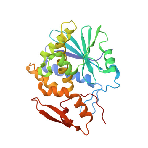
8TAB

RTA-PD00589


Experimental Data Snapshot

  • Method: X-RAY DIFFRACTION
  • Resolution: 2.26 Å
  • R-Value Free: 0.219 
  • R-Value Work: 0.186 
  • R-Value Observed: 0.187 

wwPDB Validation   3D Report Full Report


Ligand Structure Quality Assessment 


This is version 1.0 of the entry. See complete history


Literature

Structure-based design and optimization of a new class of small molecule inhibitors targeting the P-stalk binding pocket of ricin.

Rudolph, M.J.Dutta, A.Tsymbal, A.M.McLaughlin, J.E.Chen, Y.Davis, S.A.Theodorous, S.A.Pierce, M.Algava, B.Zhang, X.Szekely, Z.Roberge, J.Y.Li, X.P.Tumer, N.E.

(2024) Bioorg Med Chem 100: 117614-117614

  • DOI: https://doi.org/10.1016/j.bmc.2024.117614
  • Primary Citation of Related Structures:  
    8T9V, 8TAB, 8TAD

  • PubMed Abstract: 

    Ricin, a category-B agent for bioterrorism, and Shiga toxins (Stxs), which cause food poisoning bind to the ribosomal P-stalk to depurinate the sarcin/ricin loop. No effective therapy exists for ricin or Stx intoxication. Ribosome binding sites of the toxins have not been targeted by small molecules. We previously identified CC10501, which inhibits toxin activity by binding the P-stalk pocket of ricin toxin A subunit (RTA) remote from the catalytic site. Here, we developed a fluorescence polarization assay and identified a new class of compounds, which bind P-stalk pocket of RTA with higher affinity and inhibit catalytic activity with submicromolar potency. A lead compound, RU-NT-206, bound P-stalk pocket of RTA with similar affinity as a five-fold larger P-stalk peptide and protected cells against ricin and Stx2 holotoxins for the first time. These results validate the P-stalk binding site of RTA as a critical target for allosteric inhibition of the active site.


  • Organizational Affiliation

    New York Structural Biology Center, 89 Convent Ave, New York, NY 10027, United States.


Macromolecules
Find similar proteins by:  (by identity cutoff)  |  3D Structure
Entity ID: 1
MoleculeChains Sequence LengthOrganismDetailsImage
Ricin A chain258Ricinus communisMutation(s): 0 
EC: 3.2.2.22
UniProt
Find proteins for P02879 (Ricinus communis)
Explore P02879 
Go to UniProtKB:  P02879
Entity Groups  
Sequence Clusters30% Identity50% Identity70% Identity90% Identity95% Identity100% Identity
UniProt GroupP02879
Sequence Annotations
Expand
  • Reference Sequence
Small Molecules
Ligands 4 Unique
IDChains Name / Formula / InChI Key2D Diagram3D Interactions
2PE
Query on 2PE

Download Ideal Coordinates CCD File 
H [auth A]NONAETHYLENE GLYCOL
C18 H38 O10
YZUUTMGDONTGTN-UHFFFAOYSA-N
U4T (Subject of Investigation/LOI)
Query on U4T

Download Ideal Coordinates CCD File 
E [auth A]4H,5H-naphtho[1,2-b]thiophene-2-carboxylic acid
C13 H10 O2 S
IGBRCZHGFVMFCR-UHFFFAOYSA-N
EDO
Query on EDO

Download Ideal Coordinates CCD File 
F [auth A],
G [auth A]
1,2-ETHANEDIOL
C2 H6 O2
LYCAIKOWRPUZTN-UHFFFAOYSA-N
CL
Query on CL

Download Ideal Coordinates CCD File 
B [auth A],
C [auth A],
D [auth A]
CHLORIDE ION
Cl
VEXZGXHMUGYJMC-UHFFFAOYSA-M
Experimental Data & Validation

Experimental Data

  • Method: X-RAY DIFFRACTION
  • Resolution: 2.26 Å
  • R-Value Free: 0.219 
  • R-Value Work: 0.186 
  • R-Value Observed: 0.187 
  • Space Group: P 63 2 2
Unit Cell:
Length ( Å )Angle ( ˚ )
a = 169.146α = 90
b = 169.146β = 90
c = 55.913γ = 120
Software Package:
Software NamePurpose
PHENIXrefinement
HKL-2000data reduction
HKL-2000data scaling
PHASERphasing

Structure Validation

View Full Validation Report


Ligand Structure Quality Assessment 


Entry History & Funding Information

Deposition Data


Funding OrganizationLocationGrant Number
National Institutes of Health/National Institute Of Allergy and Infectious Diseases (NIH/NIAID)United StatesAI072425

Revision History  (Full details and data files)

  • Version 1.0: 2024-05-01
    Type: Initial release