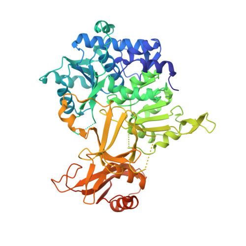
8TF7

Apo structure of protein crystal of Tri17


Experimental Data Snapshot

  • Method: X-RAY DIFFRACTION
  • Resolution: 2.45 Å
  • R-Value Free: 0.261 
  • R-Value Work: 0.216 
  • R-Value Observed: 0.218 

Starting Model: in silico
View more details

wwPDB Validation   3D Report Full Report


This is version 1.2 of the entry. See complete history


Literature

Enzymatic synthesis of azide by a promiscuous N-nitrosylase.

Del Rio Flores, A.Zhai, R.Kastner, D.W.Seshadri, K.Yang, S.De Matias, K.Shen, Y.Cai, W.Narayanamoorthy, M.Do, N.B.Xue, Z.Marzooqi, D.A.Kulik, H.J.Zhang, W.

(2024) Nat Chem 16: 2066-2075

  • DOI: https://doi.org/10.1038/s41557-024-01646-2
  • Primary Citation of Related Structures:  
    8TF7, 9BQ0

  • PubMed Abstract: 

    Azides are energy-rich compounds with diverse representation in a broad range of scientific disciplines, including material science, synthetic chemistry, pharmaceutical science and chemical biology. Despite ubiquitous usage of the azido group, the underlying biosynthetic pathways for its formation remain largely unknown. Here we report the characterization of an enzymatic route for de novo azide construction. We demonstrate that Tri17, a promiscuous ATP- and nitrite-dependent enzyme, catalyses organic azide synthesis through sequential N-nitrosation and dehydration of aryl hydrazines. Through biochemical, structural and computational analyses, we further propose a plausible molecular mechanism for azide synthesis that sets the stage for future biocatalytic applications and biosynthetic pathway engineering.


  • Organizational Affiliation

    Department of Chemical and Biomolecular Engineering, University of California Berkeley, Berkeley, CA, USA.


Macromolecules
Find similar proteins by:  (by identity cutoff)  |  3D Structure
Entity ID: 1
MoleculeChains Sequence LengthOrganismDetailsImage
AMP-binding protein
A, B
565Streptomyces tsukubensisMutation(s): 0 
Gene Names: B7R87_11280
Entity Groups  
Sequence Clusters30% Identity50% Identity70% Identity90% Identity95% Identity100% Identity
Sequence Annotations
Expand
  • Reference Sequence
Experimental Data & Validation

Experimental Data

  • Method: X-RAY DIFFRACTION
  • Resolution: 2.45 Å
  • R-Value Free: 0.261 
  • R-Value Work: 0.216 
  • R-Value Observed: 0.218 
  • Space Group: P 41 21 2
Unit Cell:
Length ( Å )Angle ( ˚ )
a = 105.992α = 90
b = 105.992β = 90
c = 235.824γ = 90
Software Package:
Software NamePurpose
PHENIXrefinement
Blu-Icedata collection
XDSdata reduction
XDSdata scaling
PHENIXphasing

Structure Validation

View Full Validation Report


Entry History & Funding Information

Deposition Data


Funding OrganizationLocationGrant Number
National Institutes of Health/National Institute of General Medical Sciences (NIH/NIGMS)United StatesR01GM136758

Revision History  (Full details and data files)

  • Version 1.0: 2024-07-10
    Type: Initial release
  • Version 1.1: 2024-10-09
    Changes: Database references, Structure summary
  • Version 1.2: 2024-12-11
    Changes: Database references