8UJI | pdb_00008uji

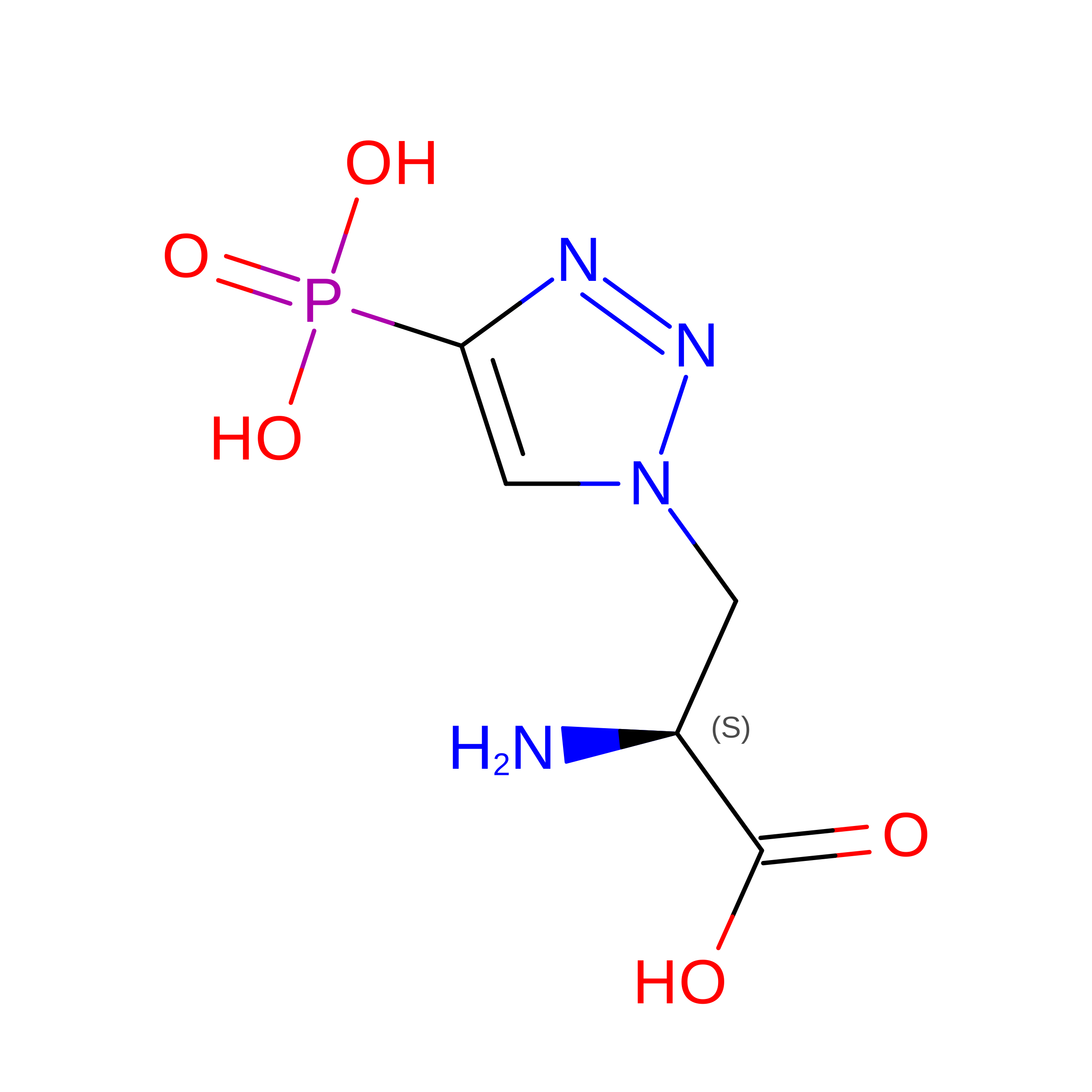
anti-Phosphohistidine Fab hSC44.ck with 3pTza peptide


Experimental Data Snapshot

  • Method: X-RAY DIFFRACTION
  • Resolution: 1.95 Å
  • R-Value Free: 
    0.260 (Depositor), 0.259 (DCC) 
  • R-Value Work: 
    0.212 (Depositor), 0.211 (DCC) 
  • R-Value Observed: 
    0.214 (Depositor) 

Starting Model: experimental
View more details

wwPDB Validation 3D Report Full Report

Validation slider image for 8UJI

This is version 1.1 of the entry. See complete history

Literature

Using phage display for rational engineering of a higher-affinity humanized 3' phosphohistidine-specific antibody.

Martyn, G.D.Kalagiri, R.Veggiani, G.Stanfield, R.L.Choudhuri, I.Sala, M.Meisenhelder, J.Chen, C.Biswas, A.Levy, R.M.Lyumkis, D.Wilson, I.A.Hunter, T.Sidhu, S.S.

(2025) Commun Chem 8: 381-381

  • DOI: https://doi.org/10.1038/s42004-025-01768-9
  • Primary Citation Related Structures: 
    8UHH, 8UHJ, 8UHN, 8UHP, 8UHS, 8UHT, 8UIG, 8UIH, 8UIO, 8UIT, 8UJI

  • PubMed Abstract: 

    Histidine phosphorylation is a non-canonical post-translational modification (PTM), with 1-phosphohistidine (1-pHis) and 3-phosphohistidine (3-pHis) isoforms, that is understudied due to a lack of robust reagents, including high-affinity pHis-specific antibodies. Engineering pHis antibodies is challenging due to the labile nature of its phosphoramidate (P-N) bond. We developed a strategy for in vitro engineering of antibodies for the detection of native 3-pHis targets, in which the rabbit SC44-8 anti-3-pTza mAb is humanized into a scaffold (hSC44) that is suitable for phage display. Six unique Fab phage-displayed hSC44 scaffold libraries were screened for antibodies that bound 3-pHis with higher affinity and had specificity for 3-pHis versus 3-pTza. hSC44.20N32F L , the best engineered antibody, has ~10-fold higher affinity for 3-pHis than parental hSC44. Eleven new Fab structures, including the first antibody-pHis peptide structures, together with structural and quantum mechanical calculations, provided molecular insights into 3-pHis and 3-pTza discrimination by hSC44.20N32F L and the increased affinity obtained through engineering. We demonstrated the utility of these high-affinity 3-pHis-specific antibodies for the recognition of pHis proteins in mammalian cells by immunoblotting and immunofluorescence staining. Our work describes a general method for engineering labile PTM-specific antibodies and provides novel antibodies for investigating the role of 3-pHis in cell biology.


  • Organizational Affiliation
    • School of Pharmacy, University of Waterloo, Kitchener, ON, Canada.

Macromolecule Content 

  • Total Structure Weight: 96.32 kDa 
  • Atom Count: 6,835 
  • Modeled Residue Count: 880 
  • Deposited Residue Count: 904 
  • Unique protein chains: 3

Macromolecules

Find similar proteins by:|  3D Structure
Entity ID: 1
MoleculeChains  Sequence LengthOrganismDetailsImage
hSC44.CK Fab heavy chainA [auth H],
C [auth A]
225Oryctolagus cuniculusMutation(s): 0 
Entity Groups
Sequence Clusters30% Identity50% Identity70% Identity90% Identity95% Identity100% Identity
Sequence Annotations
Expand
Reference Sequence
Find similar proteins by:|  3D Structure
Entity ID: 2
MoleculeChains  Sequence LengthOrganismDetailsImage
hSC44.CK Fab light chainB [auth L],
D [auth B]
218Oryctolagus cuniculusMutation(s): 0 
Entity Groups
Sequence Clusters30% Identity50% Identity70% Identity90% Identity95% Identity100% Identity
Sequence Annotations
Expand
Reference Sequence
Find similar proteins by:  Sequence   |   3D Structure  
Entity ID: 3
MoleculeChains  Sequence LengthOrganismDetailsImage
3-pTza peptideE [auth C],
F [auth D]
9synthetic constructMutation(s): 0 
Sequence Annotations
Expand
Reference Sequence

Small Molecules

Ligands 1 Unique
IDChains Name / Formula / InChI Key2D Diagram3D Interactions
EDO

Query on EDO


Download:Ideal Coordinates CCD File
G [auth H]1,2-ETHANEDIOL
C2 H6 O2
LYCAIKOWRPUZTN-UHFFFAOYSA-N

Experimental Data & Validation

Experimental Data

  • Method: X-RAY DIFFRACTION
  • Resolution: 1.95 Å
  • R-Value Free:  0.260 (Depositor), 0.259 (DCC) 
  • R-Value Work:  0.212 (Depositor), 0.211 (DCC) 
  • R-Value Observed: 0.214 (Depositor) 
Space Group: P 21 21 2
Unit Cell:
Length ( Å )Angle ( ˚ )
a = 92.498α = 90
b = 139.615β = 90
c = 73.616γ = 90
Software Package:
Software NamePurpose
PHENIXrefinement
HKL-2000data reduction
HKL-2000data scaling
PHASERphasing

Structure Validation

View Full Validation Report


Entry History 

& Funding Information

Deposition Data


Funding OrganizationLocationGrant Number
National Institutes of Health/National Cancer Institute (NIH/NCI)United StatesCA242443

Revision History  (Full details and data files)

  • Version 1.0: 2025-03-19
    Type: Initial release
  • Version 1.1: 2026-07-15
    Changes: Database references