8UQF | pdb_00008uqf

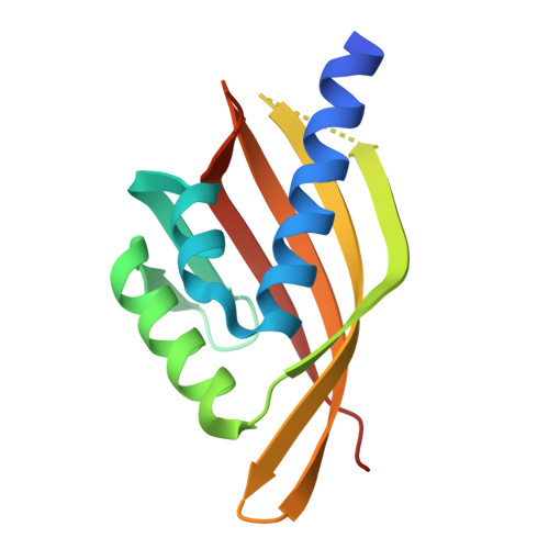
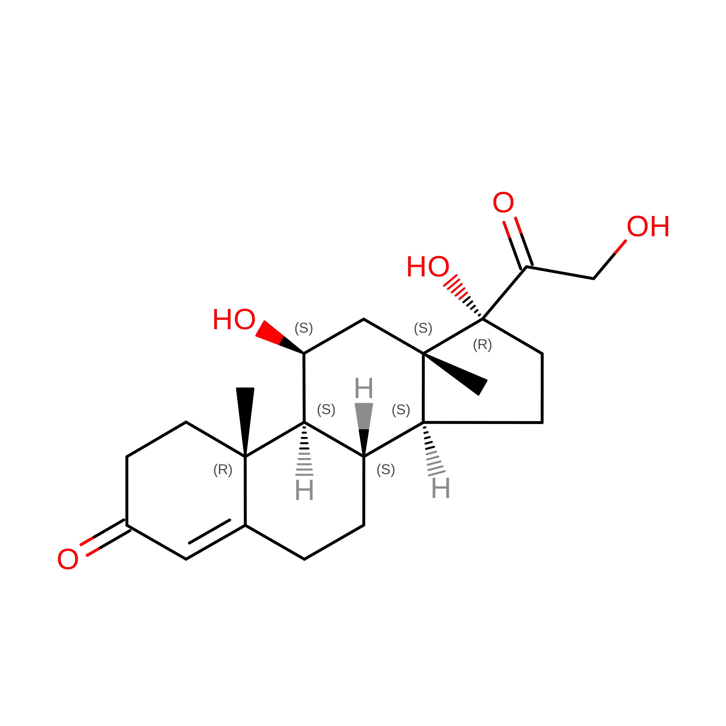
Crystal structure of designed cortisol-binding protein hcy129_mpnn5


Experimental Data Snapshot

  • Method: X-RAY DIFFRACTION
  • Resolution: 1.52 Å
  • R-Value Free: 
    0.243 (Depositor), 0.244 (DCC) 
  • R-Value Work: 
    0.227 (Depositor), 0.227 (DCC) 
  • R-Value Observed: 
    0.228 (Depositor) 

Starting Model: in silico
View more details

wwPDB Validation 3D Report Full Report

Validation slider image for 8UQF

Ligand Structure Quality Assessment 


This is version 1.1 of the entry. See complete history

Literature

Small-molecule binding and sensing with a designed protein family.

Lee, G.R.Pellock, S.J.Norn, C.Tischer, D.Dauparas, J.Anishchenko, I.Mercer, J.A.M.Kang, A.Bera, A.K.Nguyen, H.Brackenbrough, E.Sankaran, B.Goreshnik, I.Vafeados, D.Roullier, N.Han, H.L.Coventry, B.Haddox, H.K.Liu, D.R.Yeh, A.H.Baker, D.

(2026) Nat Commun 17

  • DOI: https://doi.org/10.1038/s41467-026-70953-8
  • Primary Citation Related Structures: 
    8UQF, 8VEZ, 8VFQ

  • PubMed Abstract: 

    The de novo design of small-molecule-binding proteins holds great promise as a potential tool to develop sensors on-demand for arbitrary small molecules. Here we combine deep learning and physics-based methods to generate a family of proteins with diverse and designable pocket geometries, which we employ to computationally design binders for six small-molecule targets. Biophysical characterization of the designed binders reveals nanomolar to low micromolar binding affinities and atomic-level design accuracy. Additionally, we use a cortisol binder to design a chemically induced dimerization (CID) system that enables the construction of a biosensor for cortisol detection. The approach described here demonstrates the potential of the NTF2 fold and deep learning-based protein design in sensor development, paving the way for future platforms to design binders and sensors for small molecules across analytical, environmental, and biomedical applications.


  • Organizational Affiliation
    • Department of Biochemistry, University of Washington, Seattle, WA, USA.

Macromolecule Content 

  • Total Structure Weight: 31.63 kDa 
  • Atom Count: 2,155 
  • Modeled Residue Count: 246 
  • Deposited Residue Count: 270 
  • Unique protein chains: 1

Macromolecules

Find similar proteins by:|  3D Structure
Entity ID: 1
MoleculeChains  Sequence LengthOrganismDetailsImage
designed cortisol-binding protein hcy129_mpnn5
A, B
135synthetic constructMutation(s): 0 

Experimental Data & Validation

Experimental Data

  • Method: X-RAY DIFFRACTION
  • Resolution: 1.52 Å
  • R-Value Free:  0.243 (Depositor), 0.244 (DCC) 
  • R-Value Work:  0.227 (Depositor), 0.227 (DCC) 
  • R-Value Observed: 0.228 (Depositor) 
Space Group: P 1 21 1
Unit Cell:
Length ( Å )Angle ( ˚ )
a = 48.016α = 90
b = 48.609β = 93.99
c = 56.438γ = 90
Software Package:
Software NamePurpose
PHENIXrefinement
PHENIXrefinement
XDSdata reduction
Aimlessdata scaling
PHENIXphasing

Structure Validation

View Full Validation Report


Ligand Structure Quality Assessment 


Entry History 

& Funding Information

Deposition Data


Funding OrganizationLocationGrant Number
Other private--
Howard Hughes Medical Institute (HHMI)United States--

Revision History  (Full details and data files)

  • Version 1.0: 2024-11-20
    Type: Initial release
  • Version 1.1: 2026-07-08
    Changes: Database references