8UY2 | pdb_00008uy2

Methylenetetrahydrofolate reductase from Chaetomium thermophilum DSM 1495, AdoMet-bound, Inhibited (T) State


Experimental Data Snapshot

  • Method: X-RAY DIFFRACTION
  • Resolution: 2.83 Å
  • R-Value Free: 
    0.203 (Depositor), 0.203 (DCC) 
  • R-Value Work: 
    0.187 (Depositor), 0.187 (DCC) 

Starting Model: experimental
View more details

wwPDB Validation 3D Report Full Report

Validation slider image for 8UY2

Ligand Structure Quality Assessment 


This is version 1.1 of the entry. See complete history

Literature

Structural basis of S-adenosylmethionine-dependent allosteric transition from active to inactive states in methylenetetrahydrofolate reductase.

Yamada, K.Mendoza, J.Koutmos, M.

(2024) Nat Commun 15: 5167-5167

  • DOI: https://doi.org/10.1038/s41467-024-49327-5
  • Primary Citation Related Structures: 
    8UY1, 8UY2

  • PubMed Abstract: 

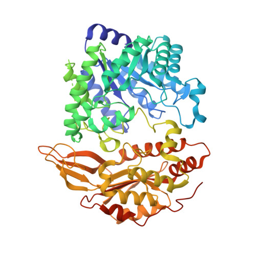
    Methylenetetrahydrofolate reductase (MTHFR) is a pivotal flavoprotein connecting the folate and methionine methyl cycles, catalyzing the conversion of methylenetetrahydrofolate to methyltetrahydrofolate. Human MTHFR (hMTHFR) undergoes elaborate allosteric regulation involving protein phosphorylation and S-adenosylmethionine (AdoMet)-dependent inhibition, though other factors such as subunit orientation and FAD status remain understudied due to the lack of a functional structural model. Here, we report crystal structures of Chaetomium thermophilum MTHFR (cMTHFR) in both active (R) and inhibited (T) states. We reveal FAD occlusion by Tyr361 in the T-state, which prevents substrate interaction. Remarkably, the inhibited form of cMTHFR accommodates two AdoMet molecules per subunit. In addition, we conducted a detailed investigation of the phosphorylation sites in hMTHFR, three of which were previously unidentified. Based on the structural framework provided by our cMTHFR model, we propose a possible mechanism to explain the allosteric structural transition of MTHFR, including the impact of phosphorylation on AdoMet-dependent inhibition.


  • Organizational Affiliation
    • Department of Chemistry, University of Michigan, Ann Arbor, MI, 48109, USA. yamadak@umich.edu.

Macromolecule Content 

  • Total Structure Weight: 288.66 kDa 
  • Atom Count: 18,670 
  • Modeled Residue Count: 2,217 
  • Deposited Residue Count: 2,468 
  • Unique protein chains: 1

Macromolecules

Find similar proteins by:|  3D Structure
Entity ID: 1
MoleculeChains  Sequence LengthOrganismDetailsImage
Methylenetetrahydrofolate reductase-like protein
A, B, C, D
617Thermochaetoides thermophila DSM 1495Mutation(s): 1 
Gene Names: CTHT_0033700
EC: 1.5.1.20
UniProt
Find proteins for G0S5U9 (Chaetomium thermophilum (strain DSM 1495 / CBS 144.50 / IMI 039719))
Explore G0S5U9 
Go to UniProtKB:  G0S5U9
Entity Groups
Sequence Clusters30% Identity50% Identity70% Identity90% Identity95% Identity100% Identity
UniProt GroupG0S5U9
Sequence Annotations
Expand
Reference Sequence

Small Molecules

Ligands 6 Unique
IDChains Name / Formula / InChI Key2D Diagram3D Interactions
FAD
(Subject of Investigation/LOI)

Query on FAD


Download:Ideal Coordinates CCD File
BA [auth D],
E [auth A],
L [auth B],
U [auth C]
FLAVIN-ADENINE DINUCLEOTIDE
C27 H33 N9 O15 P2
VWWQXMAJTJZDQX-UYBVJOGSSA-N
SAM
(Subject of Investigation/LOI)

Query on SAM


Download:Ideal Coordinates CCD File
CA [auth D]
DA [auth D]
F [auth A]
G [auth A]
M [auth B]
CA [auth D],
DA [auth D],
F [auth A],
G [auth A],
M [auth B],
N [auth B],
V [auth C],
W [auth C]
S-ADENOSYLMETHIONINE
C15 H22 N6 O5 S
MEFKEPWMEQBLKI-FCKMPRQPSA-N
SO4

Query on SO4


Download:Ideal Coordinates CCD File
AA [auth C]
EA [auth D]
FA [auth D]
J [auth A]
Q [auth B]
AA [auth C],
EA [auth D],
FA [auth D],
J [auth A],
Q [auth B],
R [auth B],
S [auth B],
T [auth B],
Z [auth C]
SULFATE ION
O4 S
QAOWNCQODCNURD-UHFFFAOYSA-L
GOL

Query on GOL


Download:Ideal Coordinates CCD File
P [auth B],
Y [auth C]
GLYCEROL
C3 H8 O3
PEDCQBHIVMGVHV-UHFFFAOYSA-N
ACT

Query on ACT


Download:Ideal Coordinates CCD File
H [auth A],
I [auth A],
O [auth B],
X [auth C]
ACETATE ION
C2 H3 O2
QTBSBXVTEAMEQO-UHFFFAOYSA-M
NA

Query on NA


Download:Ideal Coordinates CCD File
K [auth A]SODIUM ION
Na
FKNQFGJONOIPTF-UHFFFAOYSA-N

Experimental Data & Validation

Experimental Data

  • Method: X-RAY DIFFRACTION
  • Resolution: 2.83 Å
  • R-Value Free:  0.203 (Depositor), 0.203 (DCC) 
  • R-Value Work:  0.187 (Depositor), 0.187 (DCC) 
Space Group: P 2 21 21
Unit Cell:
Length ( Å )Angle ( ˚ )
a = 130.656α = 90
b = 149.948β = 90
c = 171.056γ = 90
Software Package:
Software NamePurpose
REFMACrefinement
Aimlessdata scaling
DIALSdata reduction
PHASERphasing

Structure Validation

View Full Validation Report


Ligand Structure Quality Assessment 


Entry History 

& Funding Information

Deposition Data


Funding OrganizationLocationGrant Number
National Science Foundation (NSF, United States)United States1945174

Revision History  (Full details and data files)

  • Version 1.0: 2024-06-19
    Type: Initial release
  • Version 1.1: 2024-07-03
    Changes: Database references