Download Mendeley
Structural insights into activation mechanisms on NADase of the bacterial DSR2 anti-phage defense system.
Zhang, H., Li, Y., Li, L., Chen, L., Zhu, C., Sun, L., Dong, P., Jing, D., Yang, J., Fu, L., Xiao, F., Xia, N., Li, S., Zheng, Q., Wu, Y.(2024) Sci Adv 10: eadn5691-eadn5691
- PubMed: 39083599 Search on PubMedSearch on PubMed Central
- DOI: https://doi.org/10.1126/sciadv.adn5691
- Primary Citation Related Structures:
8XEW, 8XFE, 8XFF - PubMed Abstract:
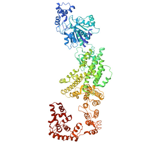
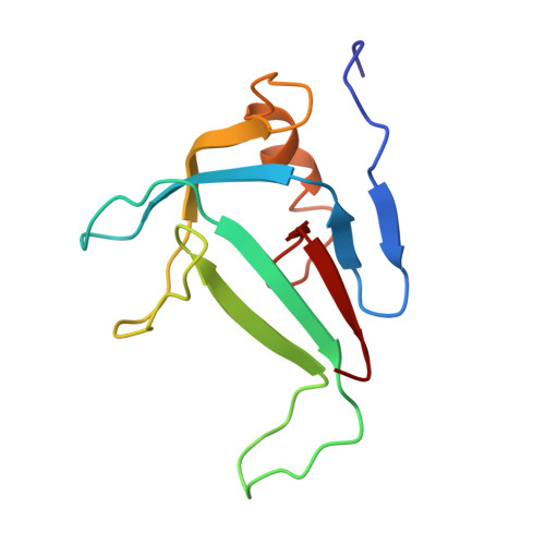
As a sirtuin (SIR2) family protein, defense-associated sirtuin2 (DSR2) has been demonstrated to participate in bacterial anti-phage resistance via depleting nicotinamide adenine dinucleotide (NAD + ) of infected cells, which can be activated by tail tube protein (TTP) and inhibited by DSR anti-defense 1 (DSAD1) of diverse phages. However, the regulating mechanism remains elusive. Here, we determined the cryo-electron microscopy structure of apo DSR2, as well as the respective complex structures with TTP and DSAD1. Structural analyses and biochemical studies reveal that DSR2 forms a tetramer with a SIR2 central core and two distinct conformations. Monomeric TTP preferentially binds to the closed conformation of DSR2, inducing conformational distortions on SIR2 tetramer assembly to activate its NADase activity. DSAD1 combines with the open conformation of DSR2, directly or allosterically inhibiting TTP activation on DSR2 NAD + hydrolysis. Our findings decipher the detailed molecule mechanisms for DSR2 NADase activity regulation and lay a foundation for in-depth understanding of the DSR2 anti-phage defense system.
- Provincial University Key Laboratory of Cellular Stress Response and Metabolic Regulation and Fujian Key Laboratory of Developmental and Neural Biology, College of Life Sciences, Fujian Normal University, Fuzhou 350117, PR China.
Organizational Affiliation: