8ZMP | pdb_00008zmp

Cryo-EM structure of the spike glycoprotein from Bat SARS-like coronavirus (Bat SL-CoV) WIV1 in locked state


Experimental Data Snapshot

  • Method: ELECTRON MICROSCOPY
  • Resolution: 3.38 Å
  • Aggregation State: PARTICLE 
  • Reconstruction Method: SINGLE PARTICLE 

Starting Model: experimental
View more details

wwPDB Validation 3D Report Full Report

Validation slider image for 8ZMP

This is version 1.2 of the entry. See complete history

Literature

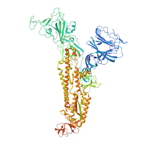
Cryo-EM structure of locked spike glycoprotein from bat SARS-like coronavirus WIV1, molecular dynamics and biophysics across host range.

Liu, C.Zheng, J.Wang, Y.Beck, F.Nagy, I.Bohn, S.Plitzko, J.M.Baumeister, W.Zhang, X.Sun, L.Zinzula, L.

(2026) Proc Natl Acad Sci U S A 123: e2516874123-e2516874123

  • DOI: https://doi.org/10.1073/pnas.2516874123
  • Primary Citation Related Structures: 
    8ZMP

  • PubMed Abstract: 

    As made evident by severe acute respiratory syndrome coronavirus 1 (SARS-CoV-1) and SARS-CoV-2 pandemics, the possibility of a SARS-like coronavirus (SL-CoV) emerging again in humankind after zoonotic spillover represents a significant global health threat. Given the role of spike (S) glycoprotein in mediating SL-CoV cell entry and cross-species transmission, there remains urgent need of structural information on SL-CoV S to guide therapeutic countermeasure development. Among SL-CoVs, bat-derived WIV1 is capable of using as receptor the angiotensin converting enzyme 2 (ACE2) from a variety of mammals, thereby representing a prototype model for studying SL-CoV precursors to future pandemics. We present a WIV1 S cryo-EM structure in prefusion state which reveals molecular signatures reminiscent of the tightly-packed locked-1 conformation described for SARS-CoV-1 and SARS-CoV-2. To decipher the molecular basis for bat SL-CoV WIV1 host range tropism, we performed molecular dynamics (MD) simulations of WIV1 S-ACE2 interaction across reservoir bat, potentially intermediate hosts civet, raccoon dog and pangolin, and accidental human hosts. We found that, in all interactions, upon complex formation with ACE2, the linoleic acid responsible for locking the S receptor binding domain (RBD) dynamically persists in its binding pocket, however repositioning to potentially unlock the system. Complex formation between WIV1 S-RBD and ACE2 from different hosts was characterized in vitro by mass photometry and microscale thermophoresis, revealing that interaction is stronger with ACE2 from bat and human than other hosts, within the latter stronger for the Thr92Ile polymorphism correlated to higher SARS-CoV-2 infection susceptibility. These findings provide critical insights with crucial implications for pandemic preparedness.


  • Organizational Affiliation
    • iHuman Institute ShanghaiTech University, Shanghai 201210, China.

Macromolecule Content 

  • Total Structure Weight: 423.34 kDa 
  • Atom Count: 26,175 
  • Modeled Residue Count: 3,198 
  • Deposited Residue Count: 3,639 
  • Unique protein chains: 1

Macromolecules

Find similar proteins by:|  3D Structure
Entity ID: 1
MoleculeChains  Sequence LengthOrganismDetailsImage
Spike glycoprotein
A, B, C
1,213Bat SARS-like coronavirus WIV1Mutation(s): 0 
UniProt
Find proteins for U5WI05 (Bat SARS-like coronavirus WIV1)
Explore U5WI05 
Go to UniProtKB:  U5WI05
Entity Groups
Sequence Clusters30% Identity50% Identity70% Identity90% Identity95% Identity100% Identity
UniProt GroupU5WI05
Glycosylation
Glycosylation Sites: 17
Sequence Annotations
Expand
Reference Sequence

Oligosaccharides

Help  
Entity ID: 3
MoleculeChains Length2D Diagram GlycosylationD Interactions
beta-D-mannopyranose-(1-4)-2-acetamido-2-deoxy-beta-D-glucopyranose-(1-4)-2-acetamido-2-deoxy-beta-D-glucopyranose
E, N, W
3N-Glycosylation
Glycosylation Resources
GlyTouCan: G15407YE
GlyCosmos: G15407YE
GlyGen: G15407YE
Entity ID: 4
MoleculeChains Length2D Diagram GlycosylationD Interactions
alpha-D-mannopyranose-(1-3)-beta-D-mannopyranose-(1-4)-2-acetamido-2-deoxy-beta-D-glucopyranose-(1-4)-2-acetamido-2-deoxy-beta-D-glucopyranose
G, P, Y
4N-Glycosylation
Glycosylation Resources
GlyTouCan: G81315DD
GlyCosmos: G81315DD
GlyGen: G81315DD

Small Molecules

Ligands 2 Unique
IDChains Name / Formula / InChI Key2D Diagram3D Interactions
EIC

Query on EIC


Download:Ideal Coordinates CCD File
EA [auth A],
NA [auth B],
WA [auth C]
LINOLEIC ACID
C18 H32 O2
OYHQOLUKZRVURQ-HZJYTTRNSA-N
NAG

Query on NAG


Download:Ideal Coordinates CCD File
AB [auth C]
BB [auth C]
CB [auth C]
DB [auth C]
EB [auth C]
AB [auth C],
BB [auth C],
CB [auth C],
DB [auth C],
EB [auth C],
FA [auth A],
GA [auth A],
HA [auth A],
IA [auth A],
JA [auth A],
KA [auth A],
LA [auth A],
MA [auth A],
OA [auth B],
PA [auth B],
QA [auth B],
RA [auth B],
SA [auth B],
TA [auth B],
UA [auth B],
VA [auth B],
XA [auth C],
YA [auth C],
ZA [auth C]
2-acetamido-2-deoxy-beta-D-glucopyranose
C8 H15 N O6
OVRNDRQMDRJTHS-FMDGEEDCSA-N

Experimental Data & Validation

Experimental Data

  • Method: ELECTRON MICROSCOPY
  • Resolution: 3.38 Å
  • Aggregation State: PARTICLE 
  • Reconstruction Method: SINGLE PARTICLE 
EM Software:
TaskSoftware PackageVersion
MODEL REFINEMENTPHENIX

Structure Validation

View Full Validation Report


Entry History 

& Funding Information

Deposition Data


Funding OrganizationLocationGrant Number
Not funded--

Revision History  (Full details and data files)

  • Version 1.0: 2025-05-28
    Type: Initial release
  • Version 1.1: 2025-07-16
    Changes: Data collection
  • Version 1.2: 2026-06-10
    Changes: Data collection, Database references