


Crystal structure of Epoxide hydrolase MdEH (Mycobacterium dioxanotrophicus)
Zhou, Y., Lan, D.M., Wang, Y.H.To be published.
Experimental Data Snapshot
Starting Model: in silico
View more details
wwPDB Validation 3D Report Full Report

Entity ID: 1 | |||||
|---|---|---|---|---|---|
| Molecule | Chains | Sequence Length | Organism | Details | Image |
| AB hydrolase-1 domain-containing protein | 293 | Mycobacterium dioxanotrophicus | Mutation(s): 0 Gene Names: BTO20_37290 | 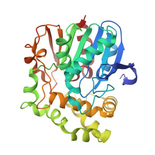 | |
UniProt | |||||
Find proteins for A0A1Y0CH10 (Mycobacterium dioxanotrophicus) Explore A0A1Y0CH10 Go to UniProtKB: A0A1Y0CH10 | |||||
Entity Groups | |||||
| Sequence Clusters | 30% Identity50% Identity70% Identity90% Identity95% Identity100% Identity | ||||
| UniProt Group | A0A1Y0CH10 | ||||
Sequence AnnotationsExpand | |||||
Reference Sequence | |||||
| Length ( Å ) | Angle ( ˚ ) |
|---|---|
| a = 179.29 | α = 90 |
| b = 179.29 | β = 90 |
| c = 151.35 | γ = 120 |
| Software Name | Purpose |
|---|---|
| PHENIX | refinement |
| HKL-3000 | data reduction |
| HKL-3000 | data scaling |
| PHENIX | phasing |
| Funding Organization | Location | Grant Number |
|---|---|---|
| Not funded | -- |


