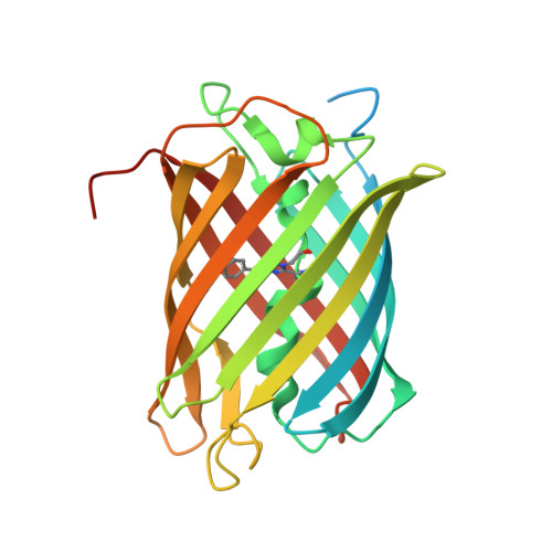
8B65 | pdb_00008b65

Structure of rsCherry crystallized in anaerobic conditions


Experimental Data Snapshot

  • Method: X-RAY DIFFRACTION
  • Resolution: 1.55 Å
  • R-Value Free: 
    0.179 (Depositor), 0.180 (DCC) 
  • R-Value Work: 
    0.151 (Depositor), 0.152 (DCC) 
  • R-Value Observed: 
    0.152 (Depositor) 

Starting Model: experimental
View more details

wwPDB Validation 3D Report Full Report

Validation slider image for 8B65

This is version 2.2 of the entry. See complete history. 

Literature

Oxygen-induced chromophore degradation in the photoswitchable red fluorescent protein rsCherry.

Bui, T.Y.H., De Zitter, E., Moeyaert, B., Pecqueur, L., Srinivasu, B.Y., Economou, A., Fontecave, M., Van Meervelt, L., Dedecker, P., Pedre, B.

(2023) Int J Biol Macromol 239: 124179-124179

  • DOI: https://doi.org/10.1016/j.ijbiomac.2023.124179
  • Primary Citation Related Structures: 
    8B65, 8B7G

  • PubMed Abstract: 

    Reversibly switchable monomeric Cherry (rsCherry) is a photoswitchable variant of the red fluorescent protein mCherry. We report that this protein gradually and irreversibly loses its red fluorescence in the dark over a period of months at 4 °C and a few days at 37 °C. We also find that its ancestor, mCherry, undergoes a similar fluorescence loss but at a slower rate. X-ray crystallography and mass spectrometry reveal that this is caused by the cleavage of the p-hydroxyphenyl ring from the chromophore and the formation of two novel types of cyclic structures at the remaining chromophore moiety. Overall, our work sheds light on a new process occurring within fluorescent proteins, further adding to the chemical diversity and versatility of these molecules.


  • Organizational Affiliation: 
    • Biochemistry, Molecular and Structural Biology Unit, Department of Chemistry, KU Leuven, Belgium.

Macromolecule Content 

  • Total Structure Weight: 31.23 kDa 
  • Atom Count: 2,222 
  • Modeled Residue Count: 221 
  • Deposited Residue Count: 270 
  • Unique protein chains: 1

Macromolecules

Find similar proteins by:|  3D Structure
Entity ID: 1
MoleculeChains  Sequence LengthOrganismDetailsImage
rsCherry269Discosoma sp.Mutation(s): 32 
UniProt
Find proteins for Q9U6Y8 (Discosoma sp.)
Explore Q9U6Y8 
Go to UniProtKB:  Q9U6Y8
Entity Groups
Sequence Clusters30% Identity50% Identity70% Identity90% Identity95% Identity100% Identity
UniProt GroupQ9U6Y8
Sequence Annotations
Expand
Reference Sequence

Small Molecules

Non-standard Monomers  2 Unique
IDChains TypeFormula2D Diagram
Q2K
Query on Q2K
L-PEPTIDE LINKINGC9 H11 N3 O4 S
QYX
Query on QYX
L-PEPTIDE LINKINGC16 H18 N3 O4 S

Experimental Data & Validation

Experimental Data

  • Method: X-RAY DIFFRACTION
  • Resolution: 1.55 Å
  • R-Value Free:  0.179 (Depositor), 0.180 (DCC) 
  • R-Value Work:  0.151 (Depositor), 0.152 (DCC) 
  • R-Value Observed: 0.152 (Depositor) 
Space Group: I 1 2 1
Unit Cell:
Length ( Å )Angle ( ˚ )
a = 61.17α = 90
b = 42.899β = 105.76
c = 108.566γ = 90
Software Package:
Software NamePurpose
XDSdata reduction
Aimlessdata scaling
PHASERphasing
PHENIXrefinement
PDB_EXTRACTdata extraction

Structure Validation

View Full Validation Report


Entry History 

& Funding Information

Deposition Data


Funding OrganizationLocationGrant Number
Other governmentViet Nam911 project
KU LeuvenBelgium--

Revision History  (Full details and data files)

  • Version 1.0: 2023-04-05
    Type: Initial release
  • Version 1.1: 2023-04-12
    Changes: Database references
  • Version 1.2: 2023-04-19
    Changes: Database references
  • Version 2.0: 2023-11-15
    Changes: Atomic model, Data collection, Derived calculations
  • Version 2.1: 2024-02-07
    Changes: Refinement description
  • Version 2.2: 2024-11-13
    Changes: Structure summary