8PCH | pdb_00008pch

CRYSTAL STRUCTURE OF PORCINE CATHEPSIN H DETERMINED AT 2.1 ANGSTROM RESOLUTION: LOCATION OF THE MINI-CHAIN C-TERMINAL CARBOXYL GROUP DEFINES CATHEPSIN H AMINOPEPTIDASE FUNCTION


Experimental Data Snapshot

  • Method: X-RAY DIFFRACTION
  • Resolution: 2.10 Å
  • R-Value Free: 
    0.245 (Depositor) 

Starting Model: experimental
View more details

wwPDB Validation 3D Report Full Report

Validation slider image for 8PCH

This is version 2.2 of the entry. See complete history

Literature

Crystal structure of porcine cathepsin H determined at 2.1 A resolution: location of the mini-chain C-terminal carboxyl group defines cathepsin H aminopeptidase function.

Guncar, G.Podobnik, M.Pungercar, J.Strukelj, B.Turk, V.Turk, D.

(1998) Structure 6: 51-61

  • DOI: https://doi.org/10.1016/s0969-2126(98)00007-0
  • Primary Citation Related Structures: 
    8PCH

  • PubMed Abstract: 

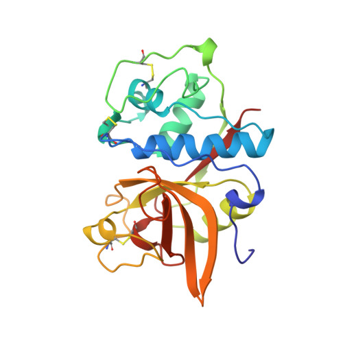
    Cathepsin H is a lysosomal cysteine protease, involved in intracellular protein degradation. It is the only known mono-aminopeptidase in the papain-like family and is reported to be involved in tumor metastasis. The cathepsin H structure was determined in order to investigate the structural basis for its aminopeptidase activity and thus to provide the basis for structure-based design of synthetic inhibitors. The crystal structure of native porcine cathepsin H was determined at 2.1 A resolution. The structure has the typical papain-family fold. The so-called mini-chain, the octapeptide EPQNCSAT, is attached via a disulfide bond to the body of the enzyme and bound in a narrowed active-site cleft, in the substrate-binding direction. The mini-chain fills the region that in related enzymes comprises the non-primed substrate-binding sites from S2 backwards. The crystal structure of cathepsin H reveals that the mini-chain has a definitive role in substrate recognition and that carbohydrate residues attached to the body of the enzyme are involved in positioning the mini-chain in the active-site cleft. Modeling of a substrate into the active-site cleft suggests that the negatively charged carboxyl group of the C terminus of the mini-chain acts as an anchor for the positively charged N-terminal amino group of a substrate. The observed displacements of the residues within the active-site cleft from their equivalent positions in the papain-like endopeptidases suggest that they form the structural basis for the positioning of both the mini-chain and the substrate, resulting in exopeptidase activity.


  • Organizational Affiliation
    • Department of Biochemistry and Molecular Biology, Jozef Stefan Institute, Ljubljana, Slovenia. gregor.guncar@ijs.si

Macromolecule Content 

  • Total Structure Weight: 25.76 kDa 
  • Atom Count: 1,982 
  • Modeled Residue Count: 228 
  • Deposited Residue Count: 228 
  • Unique protein chains: 2

Macromolecules

Find similar proteins by:|  3D Structure
Entity ID: 1
MoleculeChains  Sequence LengthOrganismDetailsImage
CATHEPSIN H220Sus scrofaMutation(s): 0 
EC: 3.4.22.16
UniProt
Find proteins for O46427 (Sus scrofa)
Explore O46427 
Go to UniProtKB:  O46427
Entity Groups
Sequence Clusters30% Identity50% Identity70% Identity90% Identity95% Identity100% Identity
UniProt GroupO46427
Glycosylation
Glycosylation Sites: 1
Sequence Annotations
Expand
Reference Sequence
Find similar proteins by:  Sequence   |   3D Structure  
Entity ID: 2
MoleculeChains  Sequence LengthOrganismDetailsImage
CATHEPSIN HB [auth P]8Sus scrofaMutation(s): 0 
EC: 3.4.22.16
UniProt
Find proteins for O46427 (Sus scrofa)
Explore O46427 
Go to UniProtKB:  O46427
Entity Groups
UniProt GroupO46427
Sequence Annotations
Expand
Reference Sequence

Oligosaccharides

Help  
Entity ID: 3
MoleculeChains Length2D Diagram GlycosylationD Interactions
beta-D-mannopyranose-(1-4)-2-acetamido-2-deoxy-beta-D-glucopyranose-(1-4)-2-acetamido-2-deoxy-beta-D-glucopyranoseC [auth B]3N-Glycosylation
Glycosylation Resources
GlyTouCan: G15407YE
GlyCosmos: G15407YE
GlyGen: G15407YE

Experimental Data & Validation

Experimental Data

  • Method: X-RAY DIFFRACTION
  • Resolution: 2.10 Å
  • R-Value Free:  0.245 (Depositor) 
Space Group: P 21 21 21
Unit Cell:
Length ( Å )Angle ( ˚ )
a = 38.442α = 90
b = 68.73β = 90
c = 86.757γ = 90
Software Package:
Software NamePurpose
DENZOdata reduction
SCALEPACKdata scaling
MAINmodel building
MAINrefinement
MAINphasing

Structure Validation

View Full Validation Report


Entry History 

Deposition Data

Revision History  (Full details and data files)

  • Version 1.0: 1998-12-09
    Type: Initial release
  • Version 1.1: 2008-03-25
    Changes: Version format compliance
  • Version 1.2: 2011-07-13
    Changes: Non-polymer description, Version format compliance
  • Version 2.0: 2020-07-29
    Type: Remediation
    Reason: Carbohydrate remediation
    Changes: Advisory, Atomic model, Data collection, Derived calculations, Structure summary
  • Version 2.1: 2023-08-09
    Changes: Advisory, Database references, Refinement description, Structure summary
  • Version 2.2: 2024-11-13
    Changes: Data collection, Structure summary