9BTC | pdb_00009btc

Cryo-EM density map of HKU1 spike glycoprotein D1 domain in complex with 9O-acetyl GD3 sialoglycan (Up state, locally refined)


Experimental Data Snapshot

  • Method: ELECTRON MICROSCOPY
  • Resolution: 2.41 Å
  • Aggregation State: PARTICLE 
  • Reconstruction Method: SINGLE PARTICLE 

wwPDB Validation   3D Report Full Report


This is version 1.1 of the entry. See complete history


Literature

Human coronavirus HKU1 spike structures reveal the basis for sialoglycan specificity and carbohydrate-promoted conformational changes.

Jin, M.Hassan, Z.Li, Z.Liu, Y.Marakhovskaia, A.Wong, A.H.M.Forman, A.Nitz, M.Gilbert, M.Yu, H.Chen, X.Rini, J.M.

(2025) Nat Commun 16: 4158-4158

  • DOI: https://doi.org/10.1038/s41467-025-59137-y
  • Primary Citation of Related Structures:  
    9BSW, 9BSX, 9BSY, 9BSZ, 9BT0, 9BT1, 9BT2, 9BT9, 9BTA, 9BTB, 9BTC, 9BTD, 9N12, 9N13, 9N14, 9N15, 9N16, 9N17, 9N18, 9N19

  • PubMed Abstract: 

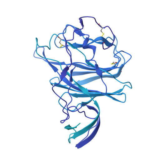
    The human coronavirus HKU1 uses both sialoglycoconjugates and the protein transmembrane serine protease 2 (TMPRSS2) as receptors. Carbohydrate binding leads to the spike protein up conformation required for TMPRSS2 binding, an outcome suggesting a distinct mechanism for driving fusion of the viral and host cell membranes. Nevertheless, the conformational changes promoted by carbohydrate binding have not been fully elucidated and the basis for HKU1's carbohydrate binding specificity remains unknown. Reported here are high resolution cryo-EM structures of the HKU1 spike protein trimer in its apo form and in complex with the carbohydrate moiety of a candidate carbohydrate receptor, the 9-O-acetylated GD3 ganglioside. The structures show that the spike monomer can exist in four discrete conformational states and that progression through them would promote the up conformation upon carbohydrate binding. We also show that a six-amino-acid insert is a determinant of HKU1's specificity for gangliosides containing a 9-O-acetylated α2-8-linked disialic acid moiety and that HKU1 shows weak affinity for the 9-O-acetylated sialic acids found on decoy receptors such as mucins.


  • Organizational Affiliation
    • Department of Biochemistry, University of Toronto, Toronto, Ontario, Canada.

Macromolecules
Find similar proteins by:  (by identity cutoff)  |  3D Structure
Entity ID: 1
MoleculeChains Sequence LengthOrganismDetailsImage
Spike glycoprotein1,194Human coronavirus HKU1Mutation(s): 0 
Gene Names: S3
UniProt
Find proteins for Q5MQD0 (Human coronavirus HKU1 (isolate N1))
Explore Q5MQD0 
Go to UniProtKB:  Q5MQD0
Entity Groups  
Sequence Clusters30% Identity50% Identity70% Identity90% Identity95% Identity100% Identity
UniProt GroupQ5MQD0
Glycosylation
Glycosylation Sites: 8
Sequence Annotations
Expand
  • Reference Sequence
Oligosaccharides

Help

Entity ID: 2
MoleculeChains Length2D Diagram Glycosylation3D Interactions
alpha-D-mannopyranose-(1-3)-[alpha-D-mannopyranose-(1-6)]beta-D-mannopyranose-(1-4)-2-acetamido-2-deoxy-beta-D-glucopyranose-(1-4)-2-acetamido-2-deoxy-beta-D-glucopyranose
B
5N-Glycosylation
Glycosylation Resources
GlyTouCan:  G22768VO
GlyCosmos:  G22768VO
GlyGen:  G22768VO
Entity ID: 3
MoleculeChains Length2D Diagram Glycosylation3D Interactions
2-acetamido-2-deoxy-beta-D-glucopyranose-(1-4)-2-acetamido-2-deoxy-beta-D-glucopyranose
C
2N-Glycosylation
Glycosylation Resources
GlyTouCan:  G42666HT
GlyCosmos:  G42666HT
GlyGen:  G42666HT
Small Molecules
Ligands 2 Unique
IDChains Name / Formula / InChI Key2D Diagram3D Interactions
A1AR1 (Subject of Investigation/LOI)
Query on A1AR1

Download Ideal Coordinates CCD File 
J [auth A]5-acetamido-8-O-(5-acetamido-9-O-acetyl-3,5-dideoxy-D-glycero-alpha-D-galacto-non-2-ulopyranonosyl)-3,5-dideoxy-D-glycero-alpha-D-galacto-non-2-ulopyranosonic acid
C24 H38 N2 O18
QVOOKWBBWNXZSQ-AWPLQUQCSA-N
NAG
Query on NAG

Download Ideal Coordinates CCD File 
D [auth A]
E [auth A]
F [auth A]
G [auth A]
H [auth A]
D [auth A],
E [auth A],
F [auth A],
G [auth A],
H [auth A],
I [auth A]
2-acetamido-2-deoxy-beta-D-glucopyranose
C8 H15 N O6
OVRNDRQMDRJTHS-FMDGEEDCSA-N
Experimental Data & Validation

Experimental Data

  • Method: ELECTRON MICROSCOPY
  • Resolution: 2.41 Å
  • Aggregation State: PARTICLE 
  • Reconstruction Method: SINGLE PARTICLE 
EM Software:
TaskSoftware PackageVersion
MODEL REFINEMENTPHENIX

Structure Validation

View Full Validation Report


Entry History & Funding Information

Deposition Data


Funding OrganizationLocationGrant Number
Canadian Institutes of Health Research (CIHR)Canada--

Revision History  (Full details and data files)

  • Version 1.0: 2025-05-14
    Type: Initial release
  • Version 1.1: 2025-05-21
    Changes: Data collection, Database references