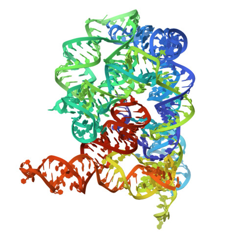
9C6J

Group IIC intron embedded with the TPP riboswitch

  • Classification: RNA
  • Organism(s): Oceanobacillus iheyensis
  • Expression System: Escherichia coli
  • Mutation(s): No 

  • Deposited: 2024-06-07 Released: 2024-12-04 
  • Deposition Author(s): Toor, N.
  • Funding Organization(s): National Institutes of Health/National Institute of General Medical Sciences (NIH/NIGMS)

Experimental Data Snapshot

  • Method: ELECTRON MICROSCOPY
  • Resolution: 2.41 Å
  • Aggregation State: PARTICLE 
  • Reconstruction Method: SINGLE PARTICLE 

wwPDB Validation   3D Report Full Report


This is version 1.0 of the entry. See complete history


Literature

Scaffold-enabled high-resolution cryo-EM structure determination of RNA.

Haack, D.B.Rudolfs, B.Jin, S.Weeks, K.M.Toor, N.

(2024) bioRxiv 

  • DOI: https://doi.org/10.1101/2024.06.10.598011
  • Primary Citation of Related Structures:  
    9C6I, 9C6J, 9C6K

  • PubMed Abstract: 

    Cryo-EM structure determination of protein-free RNAs has remained difficult with most attempts yielding low to moderate resolution and lacking nucleotide-level detail. These difficulties are compounded for small RNAs as cryo-EM is inherently more difficult for lower molecular weight macromolecules. Here we present a strategy for fusing small RNAs to a group II intron that yields high resolution structures of the appended RNA, which we demonstrate with the 86-nucleotide thiamine pyrophosphate (TPP) riboswitch, and visualizing the riboswitch ligand binding pocket at 2.5 Å resolution. We also determined the structure of the ligand-free apo state and observe that the aptamer domain of the riboswitch undergoes a large-scale conformational change upon ligand binding, illustrating how small molecule binding to an RNA can induce large effects on gene expression. This study both sets a new standard for cryo-EM riboswitch visualization and offers a versatile strategy applicable to a broad range of small to moderate-sized RNAs, which were previously intractable for high-resolution cryo-EM studies.


  • Organizational Affiliation

    Department of Chemistry and Biochemistry, University of California, San Diego, La Jolla, CA.


Macromolecules
Find similar nucleic acids by:  (by identity cutoff)  |  3D Structure
Entity ID: 1
MoleculeChains LengthOrganismImage
Group IIC intron embedded with the TPP riboswitch495Oceanobacillus iheyensis
Sequence Annotations
Expand
  • Reference Sequence
Small Molecules
Ligands 1 Unique
IDChains Name / Formula / InChI Key2D Diagram3D Interactions
MG
Query on MG

Download Ideal Coordinates CCD File 
B [auth A]
C [auth A]
D [auth A]
E [auth A]
F [auth A]
B [auth A],
C [auth A],
D [auth A],
E [auth A],
F [auth A],
G [auth A],
H [auth A],
I [auth A],
J [auth A],
K [auth A],
L [auth A],
M [auth A],
N [auth A],
O [auth A],
P [auth A],
Q [auth A],
R [auth A],
S [auth A],
T [auth A],
U [auth A],
V [auth A]
MAGNESIUM ION
Mg
JLVVSXFLKOJNIY-UHFFFAOYSA-N
Experimental Data & Validation

Experimental Data

  • Method: ELECTRON MICROSCOPY
  • Resolution: 2.41 Å
  • Aggregation State: PARTICLE 
  • Reconstruction Method: SINGLE PARTICLE 

Structure Validation

View Full Validation Report


Entry History & Funding Information

Deposition Data

  • Released Date: 2024-12-04 
  • Deposition Author(s): Toor, N.

Funding OrganizationLocationGrant Number
National Institutes of Health/National Institute of General Medical Sciences (NIH/NIGMS)United StatesR35GM141706
National Institutes of Health/National Institute of General Medical Sciences (NIH/NIGMS)United StatesR35GM122532

Revision History  (Full details and data files)

  • Version 1.0: 2024-12-04
    Type: Initial release