9CA2 | pdb_00009ca2

ENGINEERING THE HYDROPHOBIC POCKET OF CARBONIC ANHYDRASE II


Experimental Data Snapshot

  • Method: X-RAY DIFFRACTION
  • Resolution: 2.80 Å
  • R-Value Observed: 
    0.180 (Depositor) 

wwPDB Validation 3D Report Full Report

Validation slider image for 9CA2

This is version 1.4 of the entry. See complete history

Literature

Engineering the hydrophobic pocket of carbonic anhydrase II.

Alexander, R.S.Nair, S.K.Christianson, D.W.

(1991) Biochemistry 30: 11064-11072

  • DOI: https://doi.org/10.1021/bi00110a008
  • Primary Citation Related Structures: 
    4CA2, 6CA2, 7CA2, 8CA2, 9CA2

  • PubMed Abstract: 

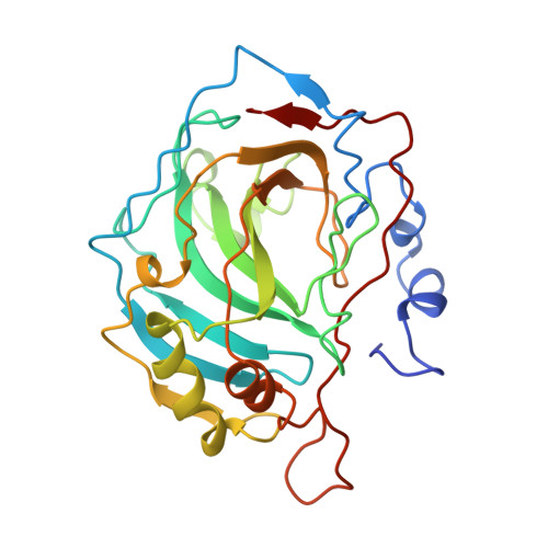
    Wild-type and mutant human carbonic anhydrases II, where mutations have been made in the hydrophobic pocket of the active site, have been studied by X-ray crystallographic methods. Specifically, mutations at Val-143 (the base of the pocket) lead to significant changes in catalytic activity and protein structure. The obliteration of a well-defined pocket in the Val-143----Phe and Val-143----Tyr mutants results in significantly diminished enzyme activity [(5 x 10(4))-fold and (3 x 10(5))-fold, respectively]; however, the activity of the Val-143----His mutant is diminished less (10(2)-fold), and deepening the pocket in the Val-143----Gly mutant results in only a 2-fold decrease in activity [Fierke et al., 1991 (preceding paper in this issue)]. These results indicate that the hydrophobic pocket is important for substrate association with the enzyme, but there are probably several catalytically acceptable substrate trajectories through this region of the enzyme structure. Additionally, each mutant protein exhibits long-range (ca. 10-15 A) compensatory structural changes which accommodate the Val-143 substitution. As such, the genetic-structural approach represented in this work serves as a three-dimensional paradigm for the redesign of specificity pockets in other protein catalysts.


  • Organizational Affiliation
    • Department of Chemistry, University of Pennsylvania, Philadelphia 19104-6323.

Macromolecule Content 

  • Total Structure Weight: 29.62 kDa 
  • Atom Count: 2,112 
  • Modeled Residue Count: 255 
  • Deposited Residue Count: 260 
  • Unique protein chains: 1

Macromolecules

Find similar proteins by:|  3D Structure
Entity ID: 1
MoleculeChains  Sequence LengthOrganismDetailsImage
CARBONIC ANHYDRASE II260Homo sapiensMutation(s): 0 
EC: 4.2.1.1 (PDB Primary Data), 4.2.1.69 (UniProt)
UniProt & NIH Common Fund Data Resources
Find proteins for P00918 (Homo sapiens)
Explore P00918 
Go to UniProtKB:  P00918
PHAROS:  P00918
GTEx:  ENSG00000104267 
Entity Groups
Sequence Clusters30% Identity50% Identity70% Identity90% Identity95% Identity100% Identity
UniProt GroupP00918
Sequence Annotations
Expand
Reference Sequence

Small Molecules

Ligands 2 Unique
IDChains Name / Formula / InChI Key2D Diagram3D Interactions
HG

Query on HG


Download:Ideal Coordinates CCD File
C [auth A]MERCURY (II) ION
Hg
BQPIGGFYSBELGY-UHFFFAOYSA-N
ZN

Query on ZN


Download:Ideal Coordinates CCD File
B [auth A]ZINC ION
Zn
PTFCDOFLOPIGGS-UHFFFAOYSA-N

Experimental Data & Validation

Experimental Data

  • Method: X-RAY DIFFRACTION
  • Resolution: 2.80 Å
  • R-Value Observed: 0.180 (Depositor) 
Space Group: P 1 21 1
Unit Cell:
Length ( Å )Angle ( ˚ )
a = 42.7α = 90
b = 41.7β = 104.6
c = 73γ = 90
Software Package:
Software NamePurpose
PROLSQrefinement

Structure Validation

View Full Validation Report


Entry History 

Deposition Data

Revision History  (Full details and data files)

  • Version 1.0: 1992-07-15
    Type: Initial release
  • Version 1.1: 2008-03-25
    Changes: Version format compliance
  • Version 1.2: 2011-07-13
    Changes: Version format compliance
  • Version 1.3: 2017-11-29
    Changes: Derived calculations, Other
  • Version 1.4: 2024-02-14
    Changes: Data collection, Database references, Derived calculations