Massively parallel assessment of designed protein solution properties using mass spectrometry and peptide barcoding.
Feldman, D., Sims, J.N., Li, X., Johnson, R., Gerben, S., Kim, D.E., Richardson, C., Koepnick, B., Eisenach, H., Hicks, D.R., Yang, E.C., Wicky, B.I.M., Milles, L.F., Bera, A.K., Kang, A., Brackenbrough, E., Joyce, E., Sankaran, B., Lubner, J.M., Goreshnik, I., Vafeados, D., Allen, A., Stewart, L., MacCoss, M.J., Baker, D.(2025) bioRxiv
- PubMed: 40060547 Search on PubMedSearch on PubMed Central
- DOI: https://doi.org/10.1101/2025.02.24.639402
- Primary Citation Related Structures:
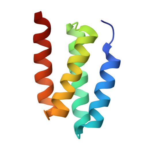
8VEA, 9DE9, 9DEA, 9DEB, 9DEC - PubMed Abstract:
Library screening and selection methods can determine the binding activities of individual members of large protein libraries given a physical link between protein and nucleotide sequence, which enables identification of functional molecules by DNA sequencing. However, the solution properties of individual protein molecules cannot be probed using such approaches because they are completely altered by DNA attachment. Mass spectrometry enables parallel evaluation of protein properties amenable to physical fractionation such as solubility and oligomeric state, but current approaches are limited to libraries of 1,000 or fewer proteins. Here, we improved mass spectrometry barcoding by co-synthesizing proteins with barcodes optimized to be highly multiplexable and minimally perturbative, scaling to libraries of >5,000 proteins. We use these barcodes together with mass spectrometry to assay the solution behavior of libraries of de novo -designed monomeric scaffolds, oligomers, binding proteins and nanocages, rapidly identifying design failure modes and successes.
- Institute for Protein Design, University of Washington, Seattle, WA 98105, USA.
Organizational Affiliation: