9EZ1

Vitamin D receptor in complex with 1,4a,25-trihydroxyvitamin D3


Experimental Data Snapshot

  • Method: X-RAY DIFFRACTION
  • Resolution: 1.95 Å
  • R-Value Free: 0.258 
  • R-Value Work: 0.208 
  • R-Value Observed: 0.212 

wwPDB Validation   3D Report Full Report


Ligand Structure Quality Assessment 


This is version 1.0 of the entry. See complete history


Literature

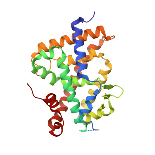
4-Hydroxy-1 alpha ,25-Dihydroxyvitamin D 3 : Synthesis and Structure-Function Study.

Peluso-Iltis, C.Pierrat, N.Rovito, D.Osz, J.Sawada, D.Kittaka, A.Laverny, G.Rochel, N.

(2024) Biomolecules 14

  • DOI: https://doi.org/10.3390/biom14050551
  • Primary Citation of Related Structures:  
    9EZ1, 9EZ2

  • PubMed Abstract: 

    The active vitamin D metabolites, 25-hydroxyvitamin D 3 (25D 3 ) and 1,25-dihydroxyvitamin D 3 (1,25D 3 ), are produced by successive hydroxylation steps and play key roles in several cellular processes. However, alternative metabolic pathways exist, and among them, the 4-hydroxylation of 25D 3 is a major one. This study aims to investigate the structure-activity relationships of 4-hydroxy derivatives of 1,25D 3 . Structural analysis indicates that 1,4α,25(OH) 3 D 3 and 1,4β,25(OH) 3 D 3 maintain the anchoring hydrogen bonds of 1,25D 3 and form additional interactions, stabilizing the active conformation of VDR. In addition, 1,4α,25D 3 and 1,4β,25D 3 are as potent as 1,25D 3 in regulating the expression of VDR target genes in rat intestinal epithelial cells and in the mouse kidney. Moreover, these two 4-hydroxy derivatives promote hypercalcemia in mice at a dose similar to that of the parent compound.


  • Organizational Affiliation

    Institute of Genetics and Molecular and Cellular Biology (IGBMC), 67400 Illkirch, France.


Macromolecules
Find similar proteins by:  (by identity cutoff)  |  3D Structure
Entity ID: 1
MoleculeChains Sequence LengthOrganismDetailsImage
Vitamin D3 receptor A302Danio rerioMutation(s): 0 
Gene Names: vdranr1i1avdr
UniProt
Find proteins for Q9PTN2 (Danio rerio)
Explore Q9PTN2 
Go to UniProtKB:  Q9PTN2
Entity Groups  
Sequence Clusters30% Identity50% Identity70% Identity90% Identity95% Identity100% Identity
UniProt GroupQ9PTN2
Sequence Annotations
Expand
  • Reference Sequence

Find similar proteins by:  Sequence   |   3D Structure  

Entity ID: 2
MoleculeChains Sequence LengthOrganismDetailsImage
Nuclear receptor coactivator 213Homo sapiensMutation(s): 0 
UniProt & NIH Common Fund Data Resources
Find proteins for Q15596 (Homo sapiens)
Explore Q15596 
Go to UniProtKB:  Q15596
PHAROS:  Q15596
GTEx:  ENSG00000140396 
Entity Groups  
Sequence Clusters30% Identity50% Identity70% Identity90% Identity95% Identity100% Identity
UniProt GroupQ15596
Sequence Annotations
Expand
  • Reference Sequence
Small Molecules
Experimental Data & Validation

Experimental Data

  • Method: X-RAY DIFFRACTION
  • Resolution: 1.95 Å
  • R-Value Free: 0.258 
  • R-Value Work: 0.208 
  • R-Value Observed: 0.212 
  • Space Group: P 65 2 2
Unit Cell:
Length ( Å )Angle ( ˚ )
a = 65.98α = 90
b = 65.98β = 90
c = 263.47γ = 120
Software Package:
Software NamePurpose
PHENIXrefinement
PHENIXrefinement
XDSdata reduction
Aimlessdata scaling
PHENIXphasing

Structure Validation

View Full Validation Report


Ligand Structure Quality Assessment 


Entry History & Funding Information

Deposition Data

  • Released Date: 2024-06-19 
  • Deposition Author(s): Rochel, N.

Funding OrganizationLocationGrant Number
Agence Nationale de la Recherche (ANR)France--

Revision History  (Full details and data files)

  • Version 1.0: 2024-06-19
    Type: Initial release