


Fine antigenic mapping of human enterovirus 71 from structures of cognate complexes for a panel of 12 patient derived monoclonal antibodies
Zhou, D., Ren, J., Stuart, D.I.To be published.
Experimental Data Snapshot
Starting Model: experimental
View more details
Entity ID: 1 | |||||
|---|---|---|---|---|---|
| Molecule | Chains | Sequence Length | Organism | Details | Image |
| heavy chain | A [auth H], C [auth A] | 234 | Homo sapiens | Mutation(s): 0 | 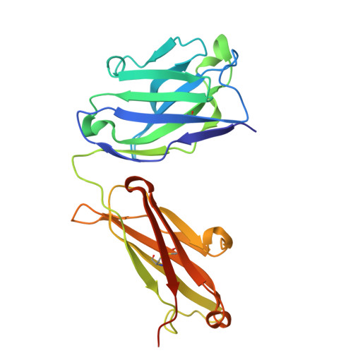 |
Entity Groups | |||||
| Sequence Clusters | 30% Identity50% Identity70% Identity90% Identity95% Identity100% Identity | ||||
Sequence AnnotationsExpand | |||||
| |||||
Entity ID: 2 | |||||
|---|---|---|---|---|---|
| Molecule | Chains | Sequence Length | Organism | Details | Image |
| light chain | B [auth L], D [auth B] | 216 | Homo sapiens | Mutation(s): 0 |  |
Entity Groups | |||||
| Sequence Clusters | 30% Identity50% Identity70% Identity90% Identity95% Identity100% Identity | ||||
Sequence AnnotationsExpand | |||||
| |||||
| Ligands 3 Unique | |||||
|---|---|---|---|---|---|
| ID | Chains | Name / Formula / InChI Key | 2D Diagram | 3D Interactions | |
| SO4 (Subject of Investigation/LOI) Query on SO4 | G [auth L], J [auth A], K [auth A], N [auth B] | SULFATE ION O4 S QAOWNCQODCNURD-UHFFFAOYSA-L |  | ||
| ACT (Subject of Investigation/LOI) Query on ACT | H [auth A], L [auth B], M [auth B] | ACETATE ION C2 H3 O2 QTBSBXVTEAMEQO-UHFFFAOYSA-M |  | ||
| CL (Subject of Investigation/LOI) Query on CL | E [auth H], F [auth L], I [auth A] | CHLORIDE ION Cl VEXZGXHMUGYJMC-UHFFFAOYSA-M |  | ||
| Length ( Å ) | Angle ( ˚ ) |
|---|---|
| a = 72.48 | α = 90 |
| b = 71.66 | β = 105.81 |
| c = 93.06 | γ = 90 |
| Software Name | Purpose |
|---|---|
| PHENIX | refinement |
| MOLREP | phasing |
| Funding Organization | Location | Grant Number |
|---|---|---|
| Medical Research Council (MRC, United Kingdom) | United Kingdom | MR/N00065X/1 |
| Wellcome Trust | United Kingdom | 090532/Z/09/Z |


