9IJX | pdb_00009ijx

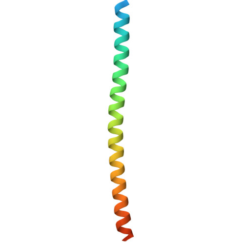
Crystal structure of the mouse Spef1 coiled-coil domain


Experimental Data Snapshot

  • Method: X-RAY DIFFRACTION
  • Resolution: 2.45 Å
  • R-Value Free: 
    0.234 (Depositor), 0.231 (DCC) 
  • R-Value Work: 
    0.195 (Depositor), 0.191 (DCC) 
  • R-Value Observed: 
    0.197 (Depositor) 

Starting Model: in silico
View more details

wwPDB Validation 3D Report Full Report

Validation slider image for 9IJX

This is version 1.1 of the entry. See complete history

Literature

Coiled-coil-mediated phase separation of Spef1 for central-pair microtubule organization and function.

Ren, J.Liu, J.Gu, K.Li, D.Qiu, B.Zhu, X.Feng, W.

(2026) Structure 34: 718-731.e6

  • DOI: https://doi.org/10.1016/j.str.2026.02.007
  • Primary Citation Related Structures: 
    9IJX

  • PubMed Abstract: 

    Central-pair microtubules (CP-MTs) are non-centrosomal MTs essential for planar beat pattern of cilia. The CP-MT formation requires the MT-associated protein Spef1, but the underlying molecular mechanism remains unclear. Here, we show that Spef1 undergoes liquid-liquid phase separation (LLPS) to facilitate non-centrosomal MT assembly by enriching tubulins. The LLPS of Spef1 is mediated by its C-terminal coiled-coil (CC) domain. Crystallography reveals that the Spef1-CC domain forms a parallel CC dimer with a unique charge distribution pattern on the surface. The dimerization capacity and charge distribution of Spef1-CC are both critical for controlling in vitro LLPS. Disruption of the dimerization capacity abolishes ciliary functions of Spef1. In contrast, a charge-changing mutant with attenuated LLPS still supports the CP-MT formation but results in cilia with abnormal beat pattern. Thus, the CC-mediated LLPS of Spef1 provides a mechanistic explanation for its prominent role in controlling CP-MT organization and function in the axoneme.


  • Organizational Affiliation
    • State Key Laboratory of Biomacromolecules, Institute of Biophysics, Chinese Academy of Sciences, 15 Datun Road, Beijing 100101, China. Electronic address: jinqi@ibp.ac.cn.

Macromolecule Content 

  • Total Structure Weight: 48.06 kDa 
  • Atom Count: 2,445 
  • Modeled Residue Count: 298 
  • Deposited Residue Count: 414 
  • Unique protein chains: 1

Macromolecules

Find similar proteins by:|  3D Structure
Entity ID: 1
MoleculeChains  Sequence LengthOrganismDetailsImage
Sperm flagellar protein 1
A, B, C, D, E
A, B, C, D, E, F
69Mus musculusMutation(s): 0 
Gene Names: Spef1Clamp
UniProt & NIH Common Fund Data Resources
Find proteins for Q99JL1 (Mus musculus)
Explore Q99JL1 
Go to UniProtKB:  Q99JL1
IMPC:  MGI:3513546
Entity Groups
Sequence Clusters30% Identity50% Identity70% Identity90% Identity95% Identity100% Identity
UniProt GroupQ99JL1
Sequence Annotations
Expand
Reference Sequence

Experimental Data & Validation

Experimental Data

  • Method: X-RAY DIFFRACTION
  • Resolution: 2.45 Å
  • R-Value Free:  0.234 (Depositor), 0.231 (DCC) 
  • R-Value Work:  0.195 (Depositor), 0.191 (DCC) 
  • R-Value Observed: 0.197 (Depositor) 
Space Group: C 1 2 1
Unit Cell:
Length ( Å )Angle ( ˚ )
a = 60.15α = 90
b = 104.36β = 90.04
c = 72.27γ = 90
Software Package:
Software NamePurpose
REFMACrefinement
XDSdata reduction
XDSdata scaling
PHASERphasing

Structure Validation

View Full Validation Report


Entry History 

& Funding Information

Deposition Data


Funding OrganizationLocationGrant Number
National Natural Science Foundation of China (NSFC)China31971160

Revision History  (Full details and data files)

  • Version 1.0: 2025-07-16
    Type: Initial release
  • Version 1.1: 2026-08-12
    Changes: Database references