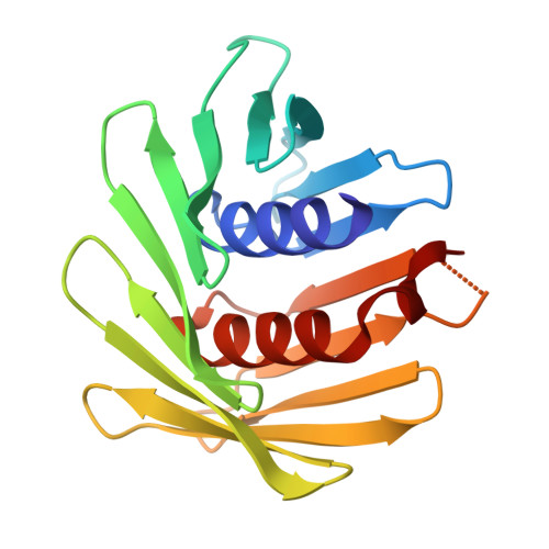
9LX4 | pdb_00009lx4

Crystal structure of the de novo designed protein ZZ4


Experimental Data Snapshot

  • Method: X-RAY DIFFRACTION
  • Resolution: 2.03 Å
  • R-Value Free: 
    0.290 (Depositor), 0.287 (DCC) 
  • R-Value Work: 
    0.223 (Depositor), 0.223 (DCC) 
  • R-Value Observed: 
    0.226 (Depositor) 

Starting Model: in silico
View more details

wwPDB Validation 3D Report Full Report

Validation slider image for 9LX4

This is version 1.2 of the entry. See complete history

Literature

Bioinformatics classification of the MgtE Mg 2 + channel and de novo protein design for the stabilization of its novel subclass.

Zhao, Z.Omae, K.Iwasaki, W.Zhang, Z.Pan, F.Lee, E.J.Ito, K.Hattori, M.

(2026) Acta Biochim Biophys Sin (Shanghai) 58: 1402-1412

  • DOI: https://doi.org/10.3724/abbs.2025224
  • Primary Citation Related Structures: 
    9LNI, 9LX4

  • PubMed Abstract: 

    MgtE channels play crucial roles in Mg 2 + homeostasis and are implicated in bacterial survival under antibiotic exposure. Previous structural and biophysical studies have focused predominantly on Thermus thermophilus MgtE, leaving the structural and mechanistic diversity of MgtE family proteins largely unexplored. In this study, via a genome mining approach, we identify diverse MgtE homologs, including a novel subclass termed the "mini-N type", which lacks the canonical cytoplasmic N and CBS domains but possesses a unique small N-like domain. Despite extensive expression screening, mini-N-type homologs cannot be stably purified. To address this issue, we design a series of de novo proteins and determine their crystal structures. A selected de novo protein is fused to a mini-N-type MgtE, enabling successful purification and preliminary cryo-EM imaging. Our findings demonstrate that de novo -designed protein fusions serve as powerful tools for stabilizing and purifying otherwise unstable membrane proteins, opening new avenues for structural and functional studies of otherwise inaccessible membrane proteins.


  • Organizational Affiliation
    • State Key Laboratory of Genetics and Development of Complex Phenotypes, Collaborative Innovation Center of Genetics and Development, Department of Physiology and Neurobiology, School of Life Sciences, Fudan University, Shanghai 200438, China.

Macromolecule Content 

  • Total Structure Weight: 38.43 kDa 
  • Atom Count: 2,708 
  • Modeled Residue Count: 350 
  • Deposited Residue Count: 354 
  • Unique protein chains: 1

Macromolecules

Find similar proteins by:|  3D Structure
Entity ID: 1
MoleculeChains  Sequence LengthOrganismDetailsImage
ZZ4
A, B
177Escherichia coliMutation(s): 0 

Small Molecules

Ligands 1 Unique
IDChains Name / Formula / InChI Key2D Diagram3D Interactions
EPE

Query on EPE


Download:Ideal Coordinates CCD File
C [auth A]4-(2-HYDROXYETHYL)-1-PIPERAZINE ETHANESULFONIC ACID
C8 H18 N2 O4 S
JKMHFZQWWAIEOD-UHFFFAOYSA-N

Experimental Data & Validation

Experimental Data

  • Method: X-RAY DIFFRACTION
  • Resolution: 2.03 Å
  • R-Value Free:  0.290 (Depositor), 0.287 (DCC) 
  • R-Value Work:  0.223 (Depositor), 0.223 (DCC) 
  • R-Value Observed: 0.226 (Depositor) 
Space Group: P 1 21 1
Unit Cell:
Length ( Å )Angle ( ˚ )
a = 57.35α = 90
b = 58.295β = 112.08
c = 67.318γ = 90
Software Package:
Software NamePurpose
PHENIXrefinement
XDSdata reduction
XDSdata scaling
PARROTphasing

Structure Validation

View Full Validation Report


Entry History 

& Funding Information

Deposition Data


Funding OrganizationLocationGrant Number
National Natural Science Foundation of China (NSFC)China--

Revision History  (Full details and data files)

  • Version 1.0: 2025-06-04
    Type: Initial release
  • Version 1.1: 2026-05-06
    Changes: Database references
  • Version 1.2: 2026-07-01
    Changes: Database references