Download Mendeley
On proline isomerization and 3D-domain-swapping.
Manjula, R., Chockalingam, N., Subramanian, R., Gosavi, S.(2026) Biochem Biophys Res Commun 821: 153872-153872
- PubMed: 42102672 Search on PubMed
- DOI: https://doi.org/10.1016/j.bbrc.2026.153872
- Primary Citation Related Structures:
6L44, 6L4I, 6L4J, 6L4N, 8TM8, 9MYK - PubMed Abstract:
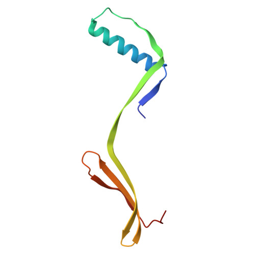
3D-domain-swapping is the exchange of identical "domains" between two protein monomers, which leads to homodimerization. This domain exchange can be facilitated by a single hinge-loop extending out. Hinge-loop prolines have been associated with domain-swapping, but their role remains unclear. Previously, we had engineered three domain-swapping variants of the monomeric monellin by replacing the wild-type hinge-loop sequence (L1:YENEGFREIKG) with QEVKG, YEIKG or QVVAG. The central residues of these sequences are hydrophobic (V/I), and lie at the apex of a tight solvent-exposed hinge-loop (L1Δ6) connecting two β-strands (β2-β3). The hydrophobic residue-solvent interaction likely impedes the closure of L1Δ6 and the formation of intra-chain β2-β3 contacts. This promotes domain-swapping. Here, we replaced the apex residue with the borderline-hydrophobic proline and found that it reduced domain-swapping in all three variants, with significant domain-swapping observed only in the most hydrophobic QVPAG construct. We then structurally characterized three monomeric (QEPKG, YEPKG, and QVPAG) and one domain-swapped dimeric (QVPAG) variants of monellin using X-ray crystallography. Interestingly, we find that the dimer has a trans-proline isomer, whereas all three monomers have a cis-proline. Thus, introducing proline into a solvent-exposed tight β-turn may be a robust method for designing cis-proline. Conversely, mutating such a naturally-occurring cis-proline to a hydrophobic amino acid may induce domain-swapping. The trans-proline in a hydrophobic hinge-loop (e.g. QVPAG), can provide rigidity to the domain-swapped dimer enabling the precise design of domain-swapping-driven protein assemblies. Overall, our mutational-design strategy is a step towards clarifying the role of prolines in 3D-domain-swapping and the rational design of proline isomerization.
- National Centre for Biological Sciences, Tata Institute of Fundamental Research, Bengaluru, 560065, India. Electronic address: manjula.iob@gmail.com.
Organizational Affiliation: