9NND | pdb_00009nnd

Structure of the HERV-K (HML-2) spike complex


Experimental Data Snapshot

  • Method: ELECTRON MICROSCOPY
  • Resolution: 2.13 Å
  • Aggregation State: PARTICLE 
  • Reconstruction Method: SINGLE PARTICLE 

wwPDB Validation   3D Report Full Report


This is version 1.1 of the entry. See complete history


Literature

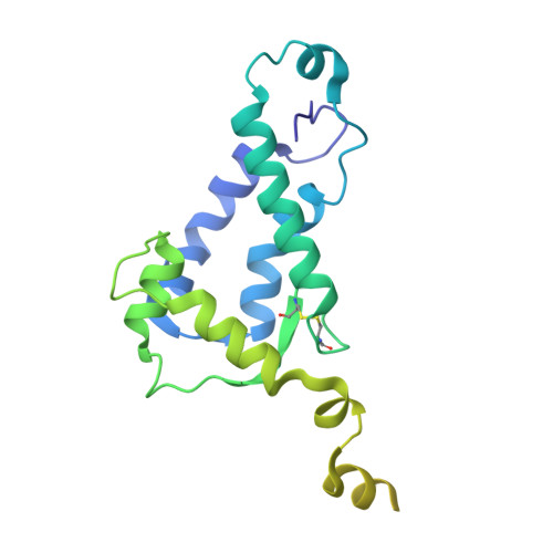
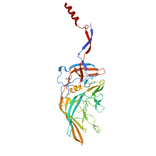
The prefusion structure of the HERV-K (HML-2) Env spike complex.

Shaked, R.Katz, M.Cohen-Dvashi, H.Diskin, R.

(2025) Proc Natl Acad Sci U S A 122: e2505505122-e2505505122

  • DOI: https://doi.org/10.1073/pnas.2505505122
  • Primary Citation of Related Structures:  
    9NND

  • PubMed Abstract: 

    The human endogenous retrovirus K (HERV-K) is a retrovirus that got assimilated into the human genome in ancient times and has been inherited in our germline ever since. It enters cells using a class-I spike protein (Env) that mediates receptor recognition and membrane fusion. On top of having a biological role during development, HERV-K is activated in amyotrophic lateral sclerosis, various cancers, and other pathological conditions. Antibodies that target the HERV-K spike complex have therapeutic value, flagging the spike as a novel drug target. Here, we use cryo-EM to determine the trimeric structure of the HERV-K spike. The spike presents a distinct structure, which substantially differs from other class-I fusogens. Nevertheless, some general architectural features suggest a common origin with other retroviruses. The ability to structurally characterize the HERV-K spike may facilitate the development of antibody-based therapies.


  • Organizational Affiliation

    Department of Chemical and Structural Biology, Weizmann Institute of Science, Rehovot 7610001, Israel.


Macromolecules
Find similar proteins by:  (by identity cutoff)  |  3D Structure
Entity ID: 1
MoleculeChains Sequence LengthOrganismDetailsImage
Endogenous retrovirus group K member 7 Pol proteinA [auth a],
C [auth c],
D [auth b]
248Homo sapiensMutation(s): 0 
Gene Names: ERVK-7
EC: 2.7.7.49 (PDB Primary Data), 3.1.26.4 (PDB Primary Data)
UniProt
Find proteins for P63135 (Homo sapiens)
Explore P63135 
Go to UniProtKB:  P63135
Entity Groups  
Sequence Clusters30% Identity50% Identity70% Identity90% Identity95% Identity100% Identity
UniProt GroupP63135
Glycosylation
Glycosylation Sites: 4Go to GlyGen: P63135-1
Sequence Annotations
Expand
  • Reference Sequence
Find similar proteins by:  (by identity cutoff)  |  3D Structure
Entity ID: 2
MoleculeChains Sequence LengthOrganismDetailsImage
Surface proteinB [auth A],
E [auth B],
F [auth C]
376Homo sapiensMutation(s): 0 
Gene Names: ERVK-25
UniProt
Find proteins for P61570 (Homo sapiens)
Explore P61570 
Go to UniProtKB:  P61570
Entity Groups  
Sequence Clusters30% Identity50% Identity70% Identity90% Identity95% Identity100% Identity
UniProt GroupP61570
Glycosylation
Glycosylation Sites: 5
Sequence Annotations
Expand
  • Reference Sequence
Oligosaccharides

Help

Entity ID: 4
MoleculeChains Length2D Diagram Glycosylation3D Interactions
2-acetamido-2-deoxy-beta-D-glucopyranose-(1-4)-[alpha-L-fucopyranose-(1-6)]2-acetamido-2-deoxy-beta-D-glucopyranoseI [auth F],
P [auth M],
S [auth P]
3N-Glycosylation
Glycosylation Resources
GlyTouCan:  G21290RB
GlyCosmos:  G21290RB
GlyGen:  G21290RB
Entity ID: 5
MoleculeChains Length2D Diagram Glycosylation3D Interactions
alpha-D-mannopyranose-(1-3)-[alpha-D-mannopyranose-(1-6)]beta-D-mannopyranose-(1-4)-2-acetamido-2-deoxy-beta-D-glucopyranose-(1-4)-2-acetamido-2-deoxy-beta-D-glucopyranoseJ [auth G],
T [auth Q],
X [auth U]
5N-Glycosylation
Glycosylation Resources
GlyTouCan:  G22768VO
GlyCosmos:  G22768VO
GlyGen:  G22768VO
Experimental Data & Validation

Experimental Data

  • Method: ELECTRON MICROSCOPY
  • Resolution: 2.13 Å
  • Aggregation State: PARTICLE 
  • Reconstruction Method: SINGLE PARTICLE 
EM Software:
TaskSoftware PackageVersion
MODEL REFINEMENTPHENIXdev_5430

Structure Validation

View Full Validation Report


Entry History & Funding Information

Deposition Data


Funding OrganizationLocationGrant Number
Not funded--

Revision History  (Full details and data files)

  • Version 1.0: 2025-07-02
    Type: Initial release
  • Version 1.1: 2025-07-16
    Changes: Data collection, Database references