


Crystal structure of AfOgg1:8OG-DNA duplex in a product-bound state
Arvai, A.S., Syed, A., Huffman, J.L., Mol, C.D., Hitomi, K., Tainer, J.A.To be published.
Experimental Data Snapshot
Starting Model: experimental
View more details
wwPDB Validation 3D Report Full Report

Entity ID: 1 | |||||
|---|---|---|---|---|---|
| Molecule | Chains | Sequence Length | Organism | Details | Image |
| 8-oxoguanine DNA glycosylase/AP lyase | 203 | Archaeoglobus fulgidus | Mutation(s): 0 Gene Names: ogg, AF_0371 EC: 3.2.2 (PDB Primary Data), 4.2.99.18 (PDB Primary Data) | 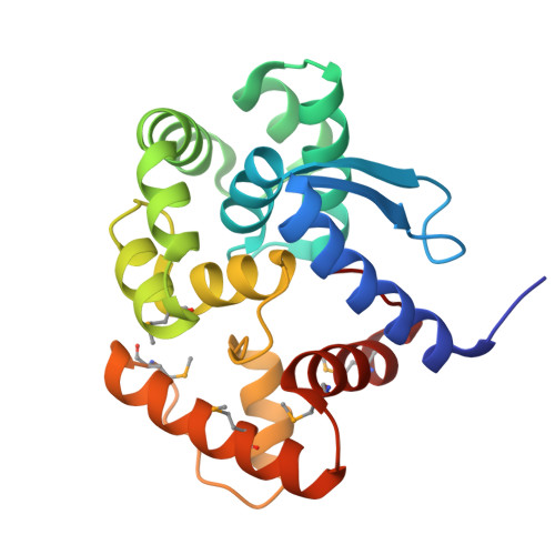 | |
UniProt | |||||
Find proteins for O29876 (Archaeoglobus fulgidus (strain ATCC 49558 / DSM 4304 / JCM 9628 / NBRC 100126 / VC-16)) Explore O29876 Go to UniProtKB: O29876 | |||||
Entity Groups | |||||
| Sequence Clusters | 30% Identity50% Identity70% Identity90% Identity95% Identity100% Identity | ||||
| UniProt Group | O29876 | ||||
Sequence AnnotationsExpand | |||||
| |||||
Find similar nucleic acids by: Sequence
Entity ID: 2 | |||||
|---|---|---|---|---|---|
| Molecule | Chains | Length | Organism | Image | |
| DNA (5'-D(P*GP*AP*GP*TP*CP(PUA))-3') | B [auth D] | 6 | synthetic construct | 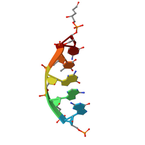 | |
Sequence AnnotationsExpand | |||||
| |||||
Find similar nucleic acids by: Sequence
Entity ID: 3 | |||||
|---|---|---|---|---|---|
| Molecule | Chains | Length | Organism | Image | |
| DNA (5'-D(P*AP*CP*CP*TP*GP*CP*A)-3') | C [auth B] | 7 | synthetic construct | 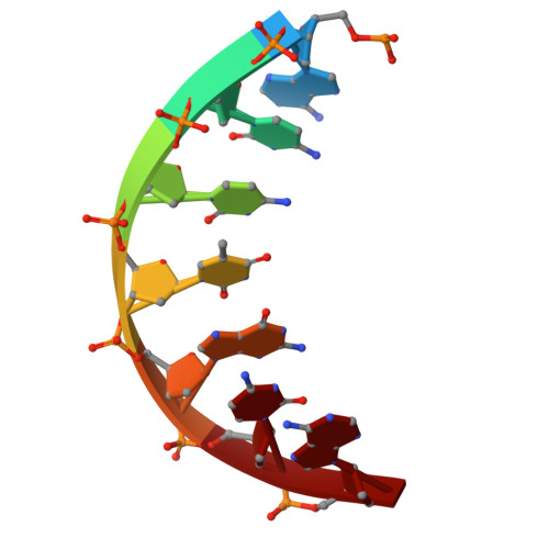 | |
Sequence AnnotationsExpand | |||||
| |||||
Find similar nucleic acids by: Sequence
Entity ID: 4 | |||||
|---|---|---|---|---|---|
| Molecule | Chains | Length | Organism | Image | |
| DNA (5'-D(*TP*GP*CP*AP*GP*GP*TP*CP*GP*AP*CP*TP*CP*TP*A)-3') | D [auth C] | 15 | synthetic construct | 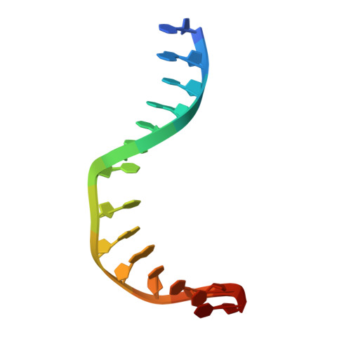 | |
Sequence AnnotationsExpand | |||||
| |||||
| Ligands 1 Unique | |||||
|---|---|---|---|---|---|
| ID | Chains | Name / Formula / InChI Key | 2D Diagram | 3D Interactions | |
| SO4 Query on SO4 | E [auth A], F [auth A] | SULFATE ION O4 S QAOWNCQODCNURD-UHFFFAOYSA-L |  | ||
| Modified Residues 1 Unique | |||||
|---|---|---|---|---|---|
| ID | Chains | Type | Formula | 2D Diagram | Parent |
| MSE Query on MSE | A | L-PEPTIDE LINKING | C5 H11 N O2 Se |  | MET |
| Length ( Å ) | Angle ( ˚ ) |
|---|---|
| a = 55.813 | α = 90 |
| b = 44.815 | β = 104.29 |
| c = 59.026 | γ = 90 |
| Software Name | Purpose |
|---|---|
| PHENIX | refinement |
| XDS | data reduction |
| XDS | data scaling |
| PHASER | phasing |
| Funding Organization | Location | Grant Number |
|---|---|---|
| National Institutes of Health/National Cancer Institute (NIH/NCI) | United States | P01 CA092584 |
| National Institutes of Health/National Cancer Institute (NIH/NCI) | United States | R35 CA220430 |


