9PC6 | pdb_00009pc6

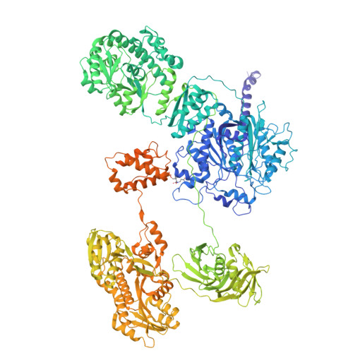
Antibody (1B2) Bound Crosslinked Rifamycin Synthetase Module 1 with a C-terminal Type II Thioesterase


Experimental Data Snapshot

  • Method: ELECTRON MICROSCOPY
  • Resolution: 3.96 Å
  • Aggregation State: PARTICLE 
  • Reconstruction Method: SINGLE PARTICLE 

Starting Model: in silico
View more details

wwPDB Validation 3D Report Full Report

Validation slider image for 9PC6

This is version 2.1 of the entry. See complete history. 

Literature

C-terminal dimerization motifs control asynchronous chain elongation during modular polyketide biosynthesis.

Liu, C., West, R.C., Chen, M., Cohn, W., Wang, G., Kim, S., Mandot, A.M., Faull, K.F., Cogan, D.P.

(2026) J Biol Chem 302: 113119-113119

  • DOI: https://doi.org/10.1016/j.jbc.2026.113119
  • Primary Citation Related Structures: 
    9PAT, 9PAV, 9PC6

  • PubMed Abstract: 

    The rifamycin synthetase (RIFS) from the bacterium Amycolatopsis mediterranei is a homodimeric assembly line that catalyzes 40+ chemical reactions to generate a complex precursor of the antitubercular drug rifampicin. It consists of an N-terminal substrate loading module followed by a decamodular polyketide synthase (PKS). While the catalytic functions are known for each domain of RIFS, how these activities are spatially and temporally coordinated during polyketide assembly remains incompletely defined. Here, we address this problem with thiol-selective crosslinking to understand the basis for conformational asymmetry during polyketide chain elongation. Our data suggest that C-terminal dimerization motifs-which are ubiquitous in bacterial PKS assembly lines-force their adjacent substrate carrier protein (CP) domains to comigrate between two equivalent ketosynthase (KS) active site chambers. Cryogenic electron microscopy analysis of the first PKS module of RIFS further underscored this observation while revealing its unique architecture. Single-turnover kinetic analysis indicated that although changes to the C-terminus that reduced CP dimerization supported 2-fold greater KS:CP interactions, they were insufficient to overcome substoichiometric product accumulation on the homodimeric protein. Our findings illuminate factors underlying asymmetry during polyketide antibiotic biosynthesis and should be instructive to future megasynth(et)ase engineers.


  • Organizational Affiliation: 
    • Department of Pharmacology and Pharmaceutical Sciences, University of Southern California, Los Angeles California, USA.

Macromolecule Content 

  • Total Structure Weight: 496.34 kDa 
  • Atom Count: 29,408 
  • Modeled Residue Count: 3,977 
  • Deposited Residue Count: 4,708 
  • Unique protein chains: 3

Macromolecules

Find similar proteins by:|  3D Structure
Entity ID: 1
MoleculeChains  Sequence LengthOrganismDetailsImage
6-deoxyerythronolide-B synthase,RifR
A, B
1,869Amycolatopsis mediterraneiMutation(s): 0 
Gene Names: rifA, rifR
EC: 2.3.1.94 (PDB Primary Data), 2.3.1.41 (UniProt)
UniProt
Find proteins for O54666 (Amycolatopsis mediterranei)
Explore O54666 
Go to UniProtKB:  O54666
Find proteins for Q7BUF9 (Amycolatopsis mediterranei)
Explore Q7BUF9 
Go to UniProtKB:  Q7BUF9
Entity Groups
Sequence Clusters30% Identity50% Identity70% Identity90% Identity95% Identity100% Identity
UniProt GroupsO54666Q7BUF9
Sequence Annotations
Expand
Reference Sequence
Find similar proteins by:|  3D Structure
Entity ID: 2
MoleculeChains  Sequence LengthOrganismDetailsImage
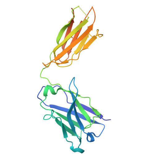
Antibody Fragment 1B2 Heavy ChainC [auth J],
D [auth K]
249Homo sapiensMutation(s): 0 
Entity Groups
Sequence Clusters30% Identity50% Identity70% Identity90% Identity95% Identity100% Identity
Sequence Annotations
Expand
Reference Sequence
Find similar proteins by:|  3D Structure
Entity ID: 3
MoleculeChains  Sequence LengthOrganismDetailsImage
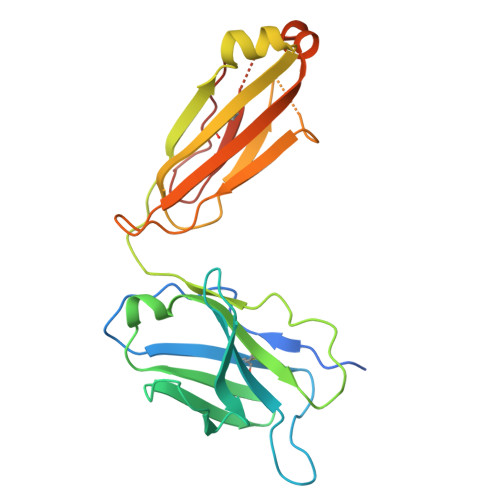
Antibody Fragment 1B2 Light ChainE [auth I],
F [auth L]
236Homo sapiensMutation(s): 0 
Entity Groups
Sequence Clusters30% Identity50% Identity70% Identity90% Identity95% Identity100% Identity
Sequence Annotations
Expand
Reference Sequence

Small Molecules

Modified Residues  1 Unique
IDChains Modified ResidueModification TypeFormula2D Diagram
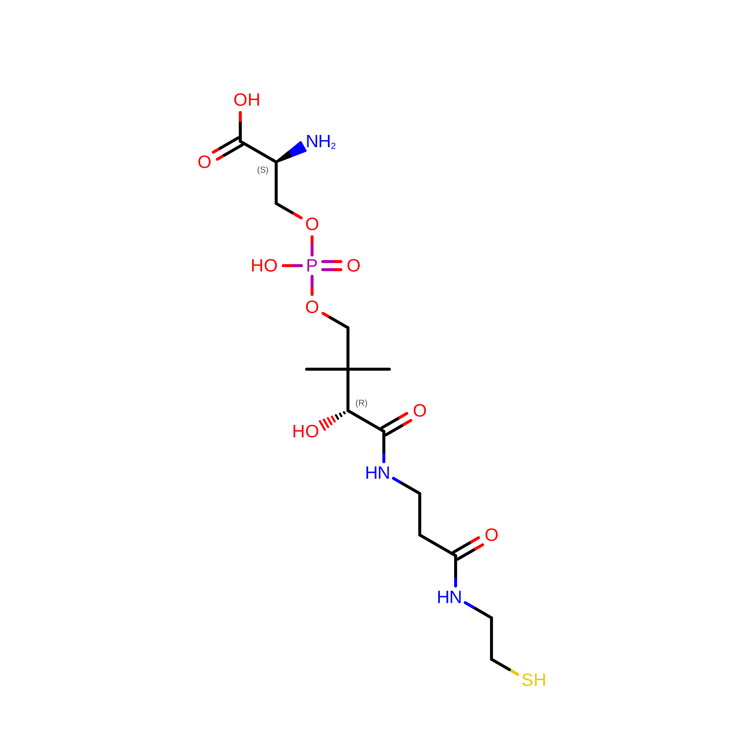
4HH
Query on 4HH
A, B
SER4-PhosphopantetheineC14 H28 N3 O9 P S

Experimental Data & Validation

Experimental Data

  • Method: ELECTRON MICROSCOPY
  • Resolution: 3.96 Å
  • Aggregation State: PARTICLE 
  • Reconstruction Method: SINGLE PARTICLE 
EM Software:
TaskSoftware PackageVersion
RECONSTRUCTIONcryoSPARC4.7.0
MODEL REFINEMENTPHENIX1.20.1_4487

Structure Validation

View Full Validation Report


Entry History 

& Funding Information

Deposition Data


Funding OrganizationLocationGrant Number
Other private--

Revision History  (Full details and data files)

  • Version 1.0: 2025-07-23
    Type: Initial release
  • Version 2.0: 2026-02-04
    Type: Coordinate replacement
    Reason: Atomic clashes
    Changes: Atomic model, Data collection, Derived calculations, Refinement description, Structure summary
  • Version 2.1: 2026-09-30
    Changes: Data collection, Database references