9R4I | pdb_00009r4i

An auto inhibitory loop in the MiDAC histone deacetylase complex


Experimental Data Snapshot

  • Method: ELECTRON MICROSCOPY
  • Resolution: 2.92 Å
  • Aggregation State: PARTICLE 
  • Reconstruction Method: SINGLE PARTICLE 

Starting Model: experimental
View more details

wwPDB Validation   3D Report Full Report


This is version 1.0 of the entry. See complete history


Literature

A de novo missense variant in MIDEAS results in increased deacetylase activity of the MiDAC HDAC complex causing a neurodevelopmental syndrome.

Fairall, L.Sirvydis, K.Turnbull, R.E.Knottnerus, S.J.Gonchar, O.Muskett, F.W.Jukes-Jones, R.van Brussel, L.van de Geer, E.van Gassen, K.Badenhorst, P.Johnson, D.Terhal, P.A.van Hasselt, P.M.van Jaarsveld, R.H.Schwabe, J.W.

(2025) Nat Commun 16: 10472-10472

  • DOI: https://doi.org/10.1038/s41467-025-65472-x
  • Primary Citation of Related Structures:  
    9R4I

  • PubMed Abstract: 

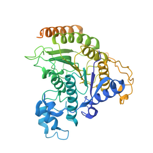
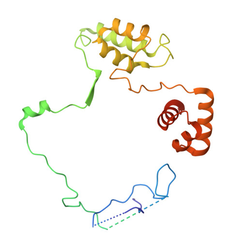
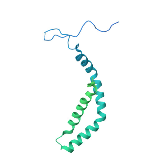
    MIDEAS is a scaffold protein that, together with DNTTIP1, mediates assembly of the MiDAC histone deacetylase complex. Mice lacking MiDAC die before birth suggesting a key developmental function. Here, we report two unrelated individuals, with a multisystem disorder characterised by delayed speech development, joint contractures, dysmorphic features and dysmotility of the gut. Both individuals have the same de novo heterozygous missense variant in MIDEAS (p.Tyr654Ser). A cryoEM structure of the MiDAC complex reveals that this amino acid is located in a conserved auto-inhibitory loop that covers the active site of the deacetylase enzyme. We suggest that the variant results in loop displacement leading to elevated deacetylase activity. In support, we observe reciprocal gene expression changes in patient fibroblasts compared with a cell line following rapid MiDAC degradation. Our results establish MIDEAS as a dominant monogenic disease gene and that hyperactivity of the MiDAC complex results in a characteristic multisystem disorder.


  • Organizational Affiliation
    • Institute for Structural and Chemical Biology, Department of Molecular and Cell Biology, University of Leicester, Leicester, UK.

Macromolecules
Find similar proteins by:  (by identity cutoff)  |  3D Structure
Entity ID: 1
MoleculeChains Sequence LengthOrganismDetailsImage
Histone deacetylase 1
A, D
482Homo sapiensMutation(s): 0 
Gene Names: HDAC1RPD3L1
EC: 3.5.1.98 (PDB Primary Data), 3.5.1 (UniProt)
UniProt & NIH Common Fund Data Resources
Find proteins for Q13547 (Homo sapiens)
Explore Q13547 
Go to UniProtKB:  Q13547
PHAROS:  Q13547
GTEx:  ENSG00000116478 
Entity Groups  
Sequence Clusters30% Identity50% Identity70% Identity90% Identity95% Identity100% Identity
UniProt GroupQ13547
Sequence Annotations
Expand
  • Reference Sequence
Find similar proteins by:  (by identity cutoff)  |  3D Structure
Entity ID: 2
MoleculeChains Sequence LengthOrganismDetailsImage
Mitotic deacetylase-associated SANT domain protein
B, E
260Homo sapiensMutation(s): 0 
Gene Names: MIDEASC14orf117C14orf43ELMSAN1
UniProt & NIH Common Fund Data Resources
Find proteins for Q6PJG2 (Homo sapiens)
Explore Q6PJG2 
Go to UniProtKB:  Q6PJG2
PHAROS:  Q6PJG2
GTEx:  ENSG00000156030 
Entity Groups  
Sequence Clusters30% Identity50% Identity70% Identity90% Identity95% Identity100% Identity
UniProt GroupQ6PJG2
Sequence Annotations
Expand
  • Reference Sequence
Find similar proteins by:  (by identity cutoff)  |  3D Structure
Entity ID: 3
MoleculeChains Sequence LengthOrganismDetailsImage
Deoxynucleotidyltransferase terminal-interacting protein 1
C, F
329Homo sapiensMutation(s): 0 
Gene Names: DNTTIP1C20orf167TDIF1
UniProt & NIH Common Fund Data Resources
Find proteins for Q9H147 (Homo sapiens)
Explore Q9H147 
Go to UniProtKB:  Q9H147
PHAROS:  Q9H147
GTEx:  ENSG00000101457 
Entity Groups  
Sequence Clusters30% Identity50% Identity70% Identity90% Identity95% Identity100% Identity
UniProt GroupQ9H147
Sequence Annotations
Expand
  • Reference Sequence
Small Molecules
Ligands 3 Unique
IDChains Name / Formula / InChI Key2D Diagram3D Interactions
IHP (Subject of Investigation/LOI)
Query on IHP

Download Ideal Coordinates CCD File 
G [auth A],
N [auth E]
INOSITOL HEXAKISPHOSPHATE
C6 H18 O24 P6
IMQLKJBTEOYOSI-GPIVLXJGSA-N
ZN
Query on ZN

Download Ideal Coordinates CCD File 
H [auth A],
K [auth D]
ZINC ION
Zn
PTFCDOFLOPIGGS-UHFFFAOYSA-N
K
Query on K

Download Ideal Coordinates CCD File 
I [auth A],
J [auth A],
L [auth D],
M [auth D]
POTASSIUM ION
K
NPYPAHLBTDXSSS-UHFFFAOYSA-N
Experimental Data & Validation

Experimental Data

  • Method: ELECTRON MICROSCOPY
  • Resolution: 2.92 Å
  • Aggregation State: PARTICLE 
  • Reconstruction Method: SINGLE PARTICLE 
EM Software:
TaskSoftware PackageVersion
MODEL REFINEMENTPHENIX1.21.1_5286
RECONSTRUCTIONcryoSPARC4.4.1

Structure Validation

View Full Validation Report


Entry History & Funding Information

Deposition Data


Funding OrganizationLocationGrant Number
Wellcome TrustUnited Kingdom222493/Z/21/Z

Revision History  (Full details and data files)

  • Version 1.0: 2026-02-25
    Type: Initial release