9R8T | pdb_00009r8t

Cryo-EM structure of the E3 ligase HECTD3


Experimental Data Snapshot

  • Method: ELECTRON MICROSCOPY
  • Resolution: 6.06 Å
  • Aggregation State: PARTICLE 
  • Reconstruction Method: SINGLE PARTICLE 

Starting Model: in silico
View more details

wwPDB Validation   3D Report Full Report


This is version 1.1 of the entry. See complete history


Literature

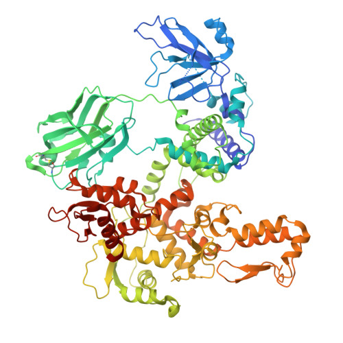
Structure and mechanism of the HECT ligase HECTD3.

Huber, J.Esposito, D.Maslen, S.Chambers, D.O.Skehel, J.M.Rittinger, K.

(2026) Nat Commun 

  • DOI: https://doi.org/10.1038/s41467-026-69520-y
  • Primary Citation of Related Structures:  
    9R6V, 9R85, 9R8T, 9R94

  • PubMed Abstract: 

    HECT E3 ligases regulate many cellular processes, yet how they recognise their substrates and synthesise specific types of poly-ubiquitin chains is still incompletely understood. HECTD3, a member of the "other HECT" family, is implicated in the regulation of inflammation, apoptosis, and infection and highly expressed in several cancers. These functions are largely attributed to its ligase activity and modification of diverse substrates with different types of ubiquitin chains. We present a detailed analysis of the ligase activity of HECTD3, including its ubiquitin linkage preferences, oligomeric state and substrate ubiquitination. Using cryo-EM, we provide the full-length structures of HECTD3 in both apo and ubiquitin-loaded forms, revealing key insights into its domain organisation, including discovery of a distinct fold of the N-terminal region, and mechanistic features. Some of these are shared with other HECT ligases, while others are unique to HECTD3 and contribute to differences in its catalytic mechanisms and functional diversity.


  • Organizational Affiliation
    • Molecular Structure of Cell Signalling Laboratory, The Francis Crick Institute, London, UK.

Macromolecules
Find similar proteins by:  (by identity cutoff)  |  3D Structure
Entity ID: 1
MoleculeChains Sequence LengthOrganismDetailsImage
E3 ubiquitin-protein ligase HECTD3864Homo sapiensMutation(s): 0 
Gene Names: HECTD3
EC: 2.3.2.26
UniProt & NIH Common Fund Data Resources
Find proteins for Q5T447 (Homo sapiens)
Explore Q5T447 
Go to UniProtKB:  Q5T447
PHAROS:  Q5T447
GTEx:  ENSG00000126107 
Entity Groups  
Sequence Clusters30% Identity50% Identity70% Identity90% Identity95% Identity100% Identity
UniProt GroupQ5T447
Sequence Annotations
Expand
  • Reference Sequence
Experimental Data & Validation

Experimental Data

  • Method: ELECTRON MICROSCOPY
  • Resolution: 6.06 Å
  • Aggregation State: PARTICLE 
  • Reconstruction Method: SINGLE PARTICLE 
EM Software:
TaskSoftware PackageVersion
RECONSTRUCTIONcryoSPARC
MODEL REFINEMENTPHENIX

Structure Validation

View Full Validation Report


Entry History & Funding Information

Deposition Data


Funding OrganizationLocationGrant Number
The Francis Crick InstituteUnited KingdomCC2075, CC2000
UK Research and Innovation (UKRI)United KingdomCC2075, CC2000
Cancer Research UKUnited KingdomCC2075, CC2000
Wellcome TrustUnited KingdomCC2075, CC2000

Revision History  (Full details and data files)

  • Version 1.0: 2026-01-28
    Type: Initial release
  • Version 1.1: 2026-03-04
    Changes: Data collection, Database references