







 Download Mendeley
Avenues to explore the selectivity of phosphoglucose isomerase inhibitors
Yan, K., Borodkin, V., Ferenbach, A., Fang, W., van Aalten, D.To be published.
Experimental Data Snapshot
Starting Model: experimental
View more details
Entity ID: 1 | |||||
|---|---|---|---|---|---|
| Molecule | Chains | Sequence Length | Organism | Details | Image |
| Glucose-6-phosphate isomerase | 555 | Candida albicans | Mutation(s): 0 Gene Names: PGI1, CAALFM_CR06340CA, CaO19.11369, CaO19.3888 EC: 5.3.1.9 (PDB Primary Data), 5.1.3 (UniProt) | 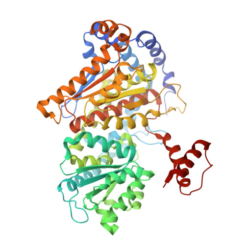 | |
UniProt | |||||
Entity Groups | |||||
| Sequence Clusters | 30% Identity50% Identity70% Identity90% Identity95% Identity100% Identity | ||||
| UniProt Group | P83780 | ||||
Sequence AnnotationsExpand | |||||
Reference Sequence | |||||
| Ligands 3 Unique | |||||
|---|---|---|---|---|---|
| ID | Chains | Name / Formula / InChI Key | 2D Diagram | 3D Interactions | |
| A1JCV (Subject of Investigation/LOI) Download:Ideal Coordinates CCD File | F [auth A], K [auth B] | [(2~{R},3~{R},4~{R},5~{S})-3,4,5-tris(oxidanyl)piperidin-2-yl]methyl dihydrogen phosphate C6 H14 N O7 P GDNPQORLESVKIU-JGWLITMVSA-N |  | ||
| IRU (Subject of Investigation/LOI) Download:Ideal Coordinates CCD File | C [auth A] D [auth A] E [auth A] G [auth A] H [auth B] | 1~{H}-pyrazol-5-ylmethanol C4 H6 N2 O UIEABCXJWANXFS-UHFFFAOYSA-N | 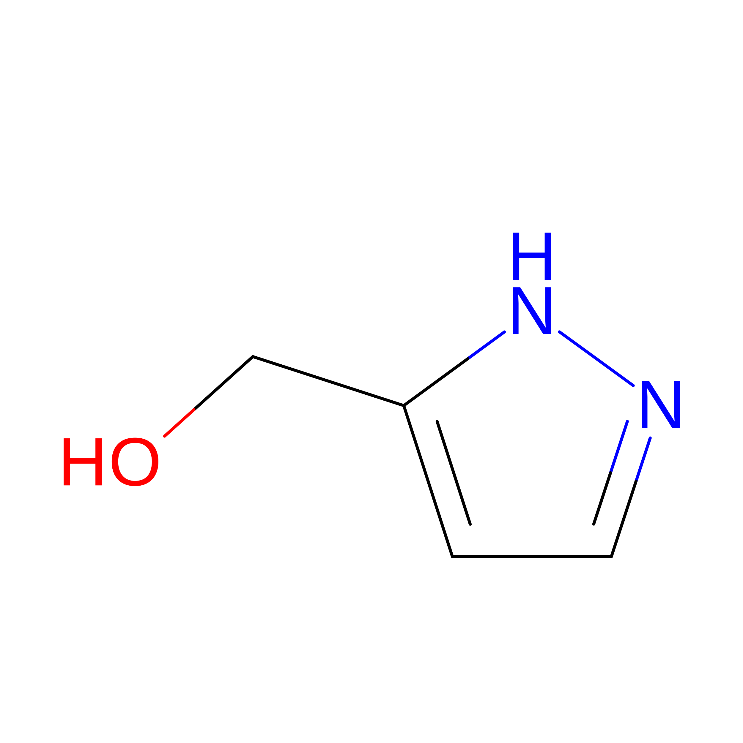 | ||
| CL Download:Ideal Coordinates CCD File | M [auth B] | CHLORIDE ION Cl VEXZGXHMUGYJMC-UHFFFAOYSA-M |  | ||
| Length ( Å ) | Angle ( ˚ ) |
|---|---|
| a = 87.37 | α = 90 |
| b = 101.499 | β = 90 |
| c = 138.913 | γ = 90 |
| Software Name | Purpose |
|---|---|
| PHENIX | refinement |
| autoPROC | data reduction |
| Aimless | data scaling |
| MOLREP | phasing |
| Funding Organization | Location | Grant Number |
|---|---|---|
| Medical Research Council (MRC, United Kingdom) | United Kingdom | MR/V001094/1 |
| Wellcome Trust | United Kingdom | 200208/Z/15/Z |








