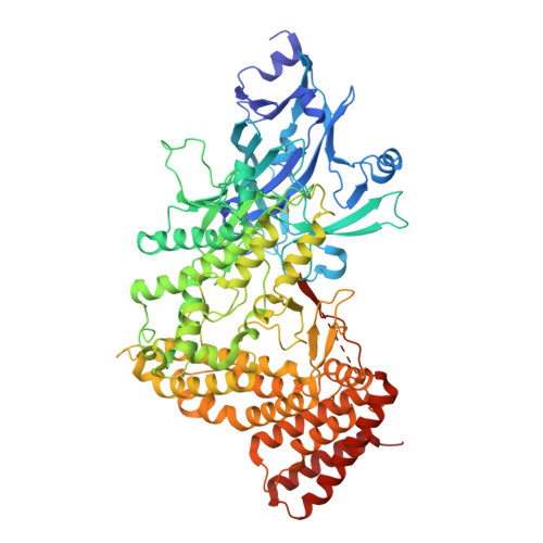
9SLL | pdb_00009sll

Zuzalysin active pentamer


Experimental Data Snapshot

  • Method: ELECTRON MICROSCOPY
  • Resolution: 2.38 Å
  • Aggregation State: PARTICLE 
  • Reconstruction Method: SINGLE PARTICLE 

wwPDB Validation 3D Report Full Report

Validation slider image for 9SLL

This is version 1.1 of the entry. See complete history. 

Literature

Structure and Function of a Multi-Megadalton Virus-Like Proteolytic Dodecahedron.

Madej, M., Rodriguez-Banqueri, A., Mizgalska, D., Szmigielski, B., Nowakowska, Z., Benedyk-Machaczka, M., Bzowska, M., Mikruta, K., Ramirez-Larrota, J.S., Agunanne, C., Julien, O., O'Donoghue, A.J., Scavenius, C., Lopez-Martin, M., Marcos, E., Koziej, L., Glatt, S., Guerra, P., Eckhard, U., Potempa, J., Gomis-Ruth, F.X.

(2026) Angew Chem Int Ed Engl : e5597279-e5597279

  • DOI: https://doi.org/10.1002/anie.5597279
  • Primary Citation Related Structures: 
    9SLL, 9SLN, 9SM4, 9SM8, 9SMJ

  • PubMed Abstract: 

    Natural pentamer dodecahedra (Ddhs) span six orders of magnitude in diameter. Among proteins, only two catalytic Ddhs have been structurally characterized: lumazine synthase (LS) and the core of pyruvate dehydrogenase (PDH). Zuzalysin (ZUZ) is a ≈95-kDa metallopeptidase secreted for virulence by Porphyromonas gingivalis. Calcium converts latent flexible monomers into active ≈0.5-MDa pentamers that further assemble hierarchically into bipentamers, tripentamers, and a ≈5.6-MDa, ≈355-Å virus-like dodecahedron (Ddh ZUZ ). Experimental structures (1.8-3.6 Å) across these states reveal the molecular basis of activation, association, and catalysis, culminating in Ddh ZUZ , which is physiologic, exceeds small viral capsids, and has 20 main entry pores and 60 lumen-facing active sites. ZUZ represents the largest catalytic protein assembly resolved at high resolution, exceeding LS, PDH, and major peptidase complexes in size and/or resolution.


  • Organizational Affiliation: 
    • Department of Microbiology, Faculty of Biochemistry, Biophysics and Biotechnology, Jagiellonian University, Kraków, Poland.

Macromolecule Content 

  • Total Structure Weight: 474.19 kDa 
  • Atom Count: 34,563 
  • Modeled Residue Count: 3,880 
  • Deposited Residue Count: 4,140 
  • Unique protein chains: 1

Macromolecules

Find similar proteins by:|  3D Structure
Entity ID: 1
MoleculeChains  Sequence LengthOrganismDetailsImage
Zinc-dependent metalloprotease
A, B, C, D, E
828Porphyromonas gingivalisMutation(s): 0 
Gene Names: NY149_10785
UniProt
Find proteins for A0AAF0BD41 (Porphyromonas gingivalis)
Explore A0AAF0BD41 
Go to UniProtKB:  A0AAF0BD41
Entity Groups
Sequence Clusters30% Identity50% Identity70% Identity90% Identity95% Identity100% Identity
UniProt GroupA0AAF0BD41
Sequence Annotations
Expand
Reference Sequence

Small Molecules

Ligands 2 Unique
IDChains Name / Formula / InChI Key2D Diagram3D Interactions
ZN
(Subject of Investigation/LOI)

Query on ZN


Download:Ideal Coordinates CCD File
H [auth A],
K [auth B],
N [auth C],
Q [auth D],
T [auth E]
ZINC ION
Zn
PTFCDOFLOPIGGS-UHFFFAOYSA-N
CA
(Subject of Investigation/LOI)

Query on CA


Download:Ideal Coordinates CCD File
F [auth A]
G [auth A]
I [auth B]
J [auth B]
L [auth C]
F [auth A],
G [auth A],
I [auth B],
J [auth B],
L [auth C],
M [auth C],
O [auth D],
P [auth D],
R [auth E],
S [auth E]
CALCIUM ION
Ca
BHPQYMZQTOCNFJ-UHFFFAOYSA-N

Experimental Data & Validation

Experimental Data

  • Method: ELECTRON MICROSCOPY
  • Resolution: 2.38 Å
  • Aggregation State: PARTICLE 
  • Reconstruction Method: SINGLE PARTICLE 
EM Software:
TaskSoftware PackageVersion
MODEL REFINEMENTPHENIX2.0rc1_5617
RECONSTRUCTIONcryoSPARC

Structure Validation

View Full Validation Report


Entry History 

& Funding Information

Deposition Data


Funding OrganizationLocationGrant Number
Not funded--

Revision History  (Full details and data files)

  • Version 1.0: 2026-09-16
    Type: Initial release
  • Version 1.1: 2026-09-23
    Changes: Data collection, Database references