9SOE | pdb_00009soe

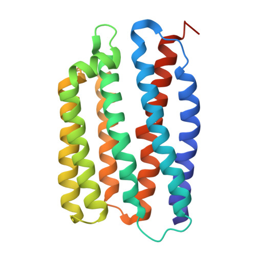
Crystal structure of viral channelrhodopsin OLPVR1 in a complex with barium ion bound at the surface


Experimental Data Snapshot

  • Method: X-RAY DIFFRACTION
  • Resolution: 2.10 Å
  • R-Value Free: 
    0.249 (Depositor) 
  • R-Value Work: 
    0.207 (Depositor) 
  • R-Value Observed: 
    0.209 (Depositor) 

Starting Model: experimental
View more details

wwPDB Validation

Currently 9SOE does not have a validation slider image.


This is version 1.1 of the entry. See complete history. 

Literature

Molecular mechanism of calcium inhibition in viral channelrhodopsins.

Zabelskii, D., Bukhdruker, S., Lamm, G.H.U., Bukhalovich, S., Aoyama, M., Sudarev, V., Kuzmin, A., Shibata, M., Kotayama, K., Kandori, H., Wachtveitl, J., Bamberg, E., Gordeliy, V.

(2026) Nat Commun 17

  • DOI: https://doi.org/10.1038/s41467-026-77716-5
  • Primary Citation Related Structures: 
    9SOD, 9SOE, 9SOF, 9SOG

  • PubMed Abstract: 

    Viral channelrhodopsins (VCR1s) are giant-virus-encoded light-gated channels permeable to monovalent and divalent cations, including Na + and Ca 2+ ions, and inhibited by millimolar Ca 2+ concentrations. Here, we combine X-ray crystallography, time-resolved UV-vis spectroscopy, and ATR-FTIR spectroscopy to investigate molecular mechanisms of ion permeation and Ca 2+ -dependent inhibition in OLPVR1. An atomic resolution structure of OLPVR1 obtained in the presence of 10 mM CaCl 2 and 900 mM NaCl reveals a transient intracellular Ca 2+ binding site near T87 and T88, close to the retinal cofactor. Upon photoactivation, this Ca 2+ ion prevents a key rearrangement of the intracellular gate required for ion translocation, namely the flip of E44, thereby disrupting ion conduction. Instead, illumination leads to the accumulation of Na + ions between E44, S208 and the carbonyl oxygen of retinal-binding residue K204. Our findings reveal the molecular basis of Ca 2+ -dependent inhibition in VCR1s and provide a foundation for engineering enhanced tools for calcium optogenetics.


  • Organizational Affiliation: 
    • European X-ray Free Electron Laser GmbH, Schenefeld, Germany. dmitrii.zabelskii@xfel.eu.

Macromolecule Content 

  • Total Structure Weight: 34.41 kDa 
  • Atom Count: 2,233 
  • Modeled Residue Count: 223 
  • Deposited Residue Count: 231 
  • Unique protein chains: 1

Macromolecules

Find similar proteins by:|  3D Structure
Entity ID: 1
MoleculeChains  Sequence LengthOrganismDetailsImage
rhodopsin231Organic Lake phycodnavirusMutation(s): 0 
Gene Names: 162281038
Entity Groups
Sequence Clusters30% Identity50% Identity70% Identity90% Identity95% Identity100% Identity
Sequence Annotations
Expand
Reference Sequence

Small Molecules

Ligands 7 Unique
IDChains Name / Formula / InChI Key2D Diagram3D Interactions
OLC

Query on OLC


Download:Ideal Coordinates CCD File
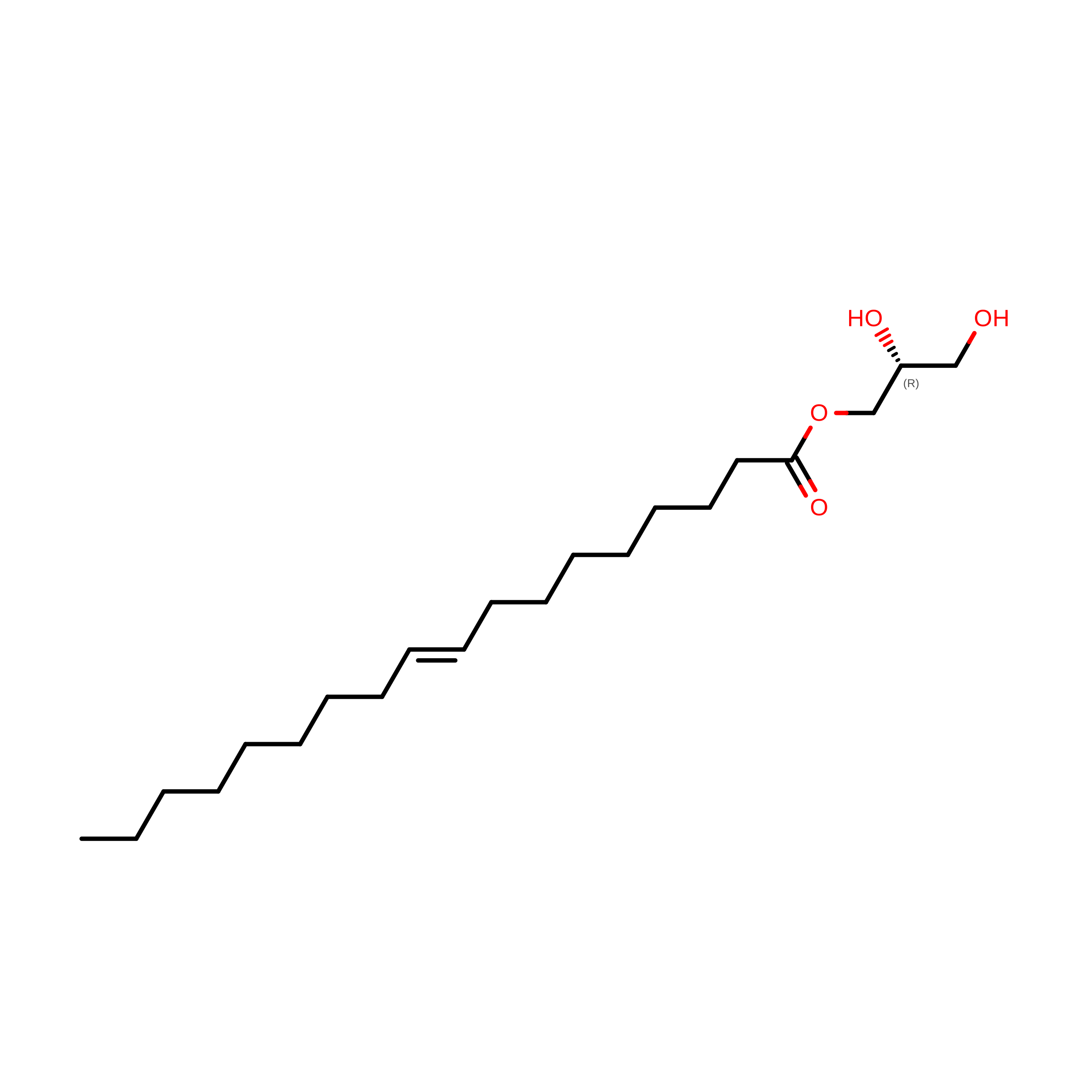
U [auth A](2R)-2,3-dihydroxypropyl (9Z)-octadec-9-enoate
C21 H40 O4
RZRNAYUHWVFMIP-GDCKJWNLSA-N
97N

Query on 97N


Download:Ideal Coordinates CCD File
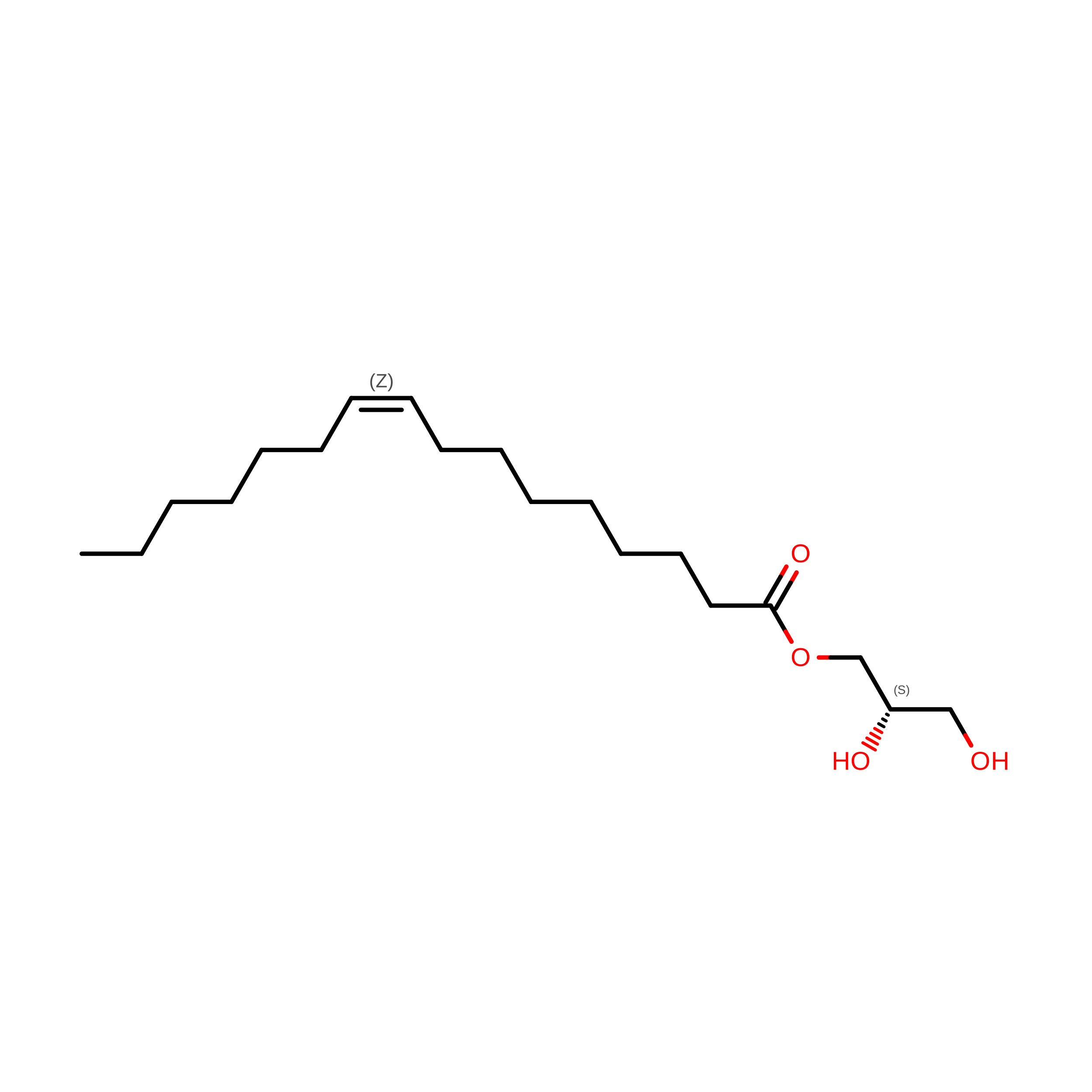
R [auth A](2S)-2,3-dihydroxypropyl (9Z)-hexadec-9-enoate
C19 H36 O4
KVYUBFKSKZWZSV-ZEVQVBBLSA-N
RET

Query on RET


Download:Ideal Coordinates CCD File
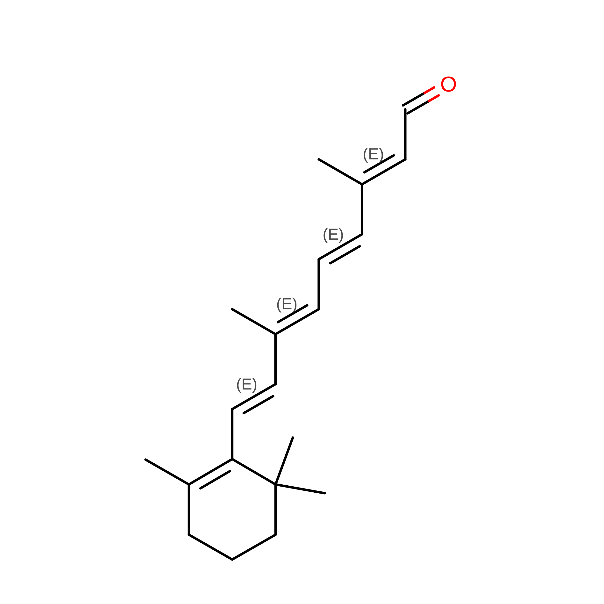
Y [auth A]RETINAL
C20 H28 O
NCYCYZXNIZJOKI-OVSJKPMPSA-N
LFA

Query on LFA


Download:Ideal Coordinates CCD File
B [auth A]
C [auth A]
D [auth A]
E [auth A]
F [auth A]
B [auth A],
C [auth A],
D [auth A],
E [auth A],
F [auth A],
G [auth A],
H [auth A],
I [auth A],
J [auth A],
K [auth A],
L [auth A],
M [auth A],
N [auth A],
O [auth A],
P [auth A],
Q [auth A],
S [auth A],
V [auth A]
EICOSANE
C20 H42
CBFCDTFDPHXCNY-UHFFFAOYSA-N
BA(
Subject of Investigation/LOI)

Query on BA


Download:Ideal Coordinates CCD File
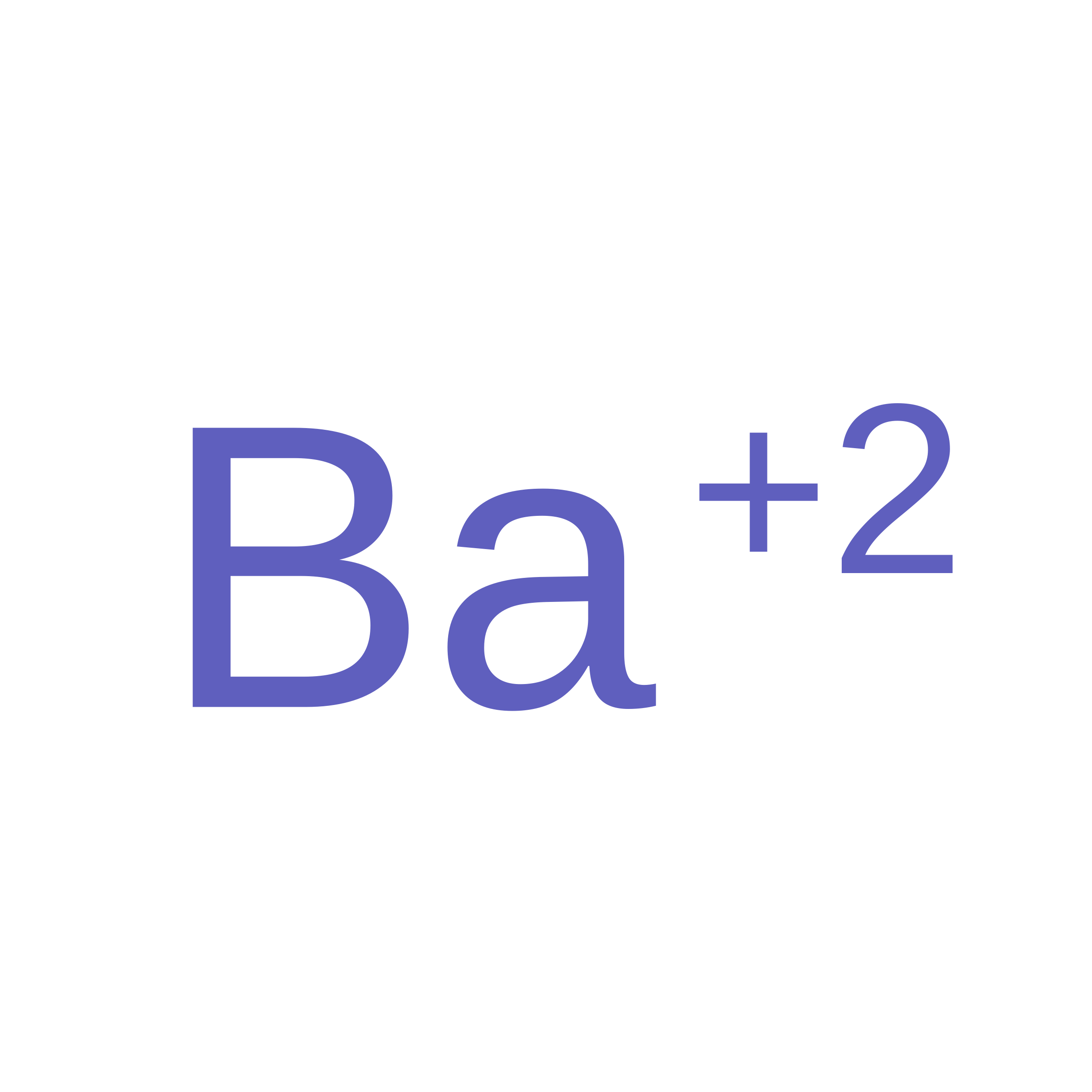
X [auth A]BARIUM ION
Ba
XDFCIPNJCBUZJN-UHFFFAOYSA-N
GOL

Query on GOL


Download:Ideal Coordinates CCD File
T [auth A]GLYCEROL
C3 H8 O3
PEDCQBHIVMGVHV-UHFFFAOYSA-N
NA

Query on NA


Download:Ideal Coordinates CCD File
W [auth A]SODIUM ION
Na
FKNQFGJONOIPTF-UHFFFAOYSA-N

Experimental Data & Validation

Experimental Data

  • Method: X-RAY DIFFRACTION
  • Resolution: 2.10 Å
  • R-Value Free:  0.249 (Depositor) 
  • R-Value Work:  0.207 (Depositor) 
  • R-Value Observed: 0.209 (Depositor) 
Space Group: P 21 21 2
Unit Cell:
Length ( Å )Angle ( ˚ )
a = 46.219α = 90
b = 115.034β = 90
c = 53.294γ = 90
Software Package:
Software NamePurpose
XDSdata reduction
XSCALEdata scaling
PHASERphasing
PHENIXrefinement

Structure Validation

Currently 9SOE does not have a validation slider image.


Entry History 

& Funding Information

Deposition Data


Funding OrganizationLocationGrant Number
Agence Nationale de la Recherche (ANR)FranceANR-19-CE11-0026

Revision History  (Full details and data files)

  • Version 1.0: 2026-09-23
    Type: Initial release
  • Version 1.1: 2026-09-30
    Changes: Database references