9T1O | pdb_00009t1o

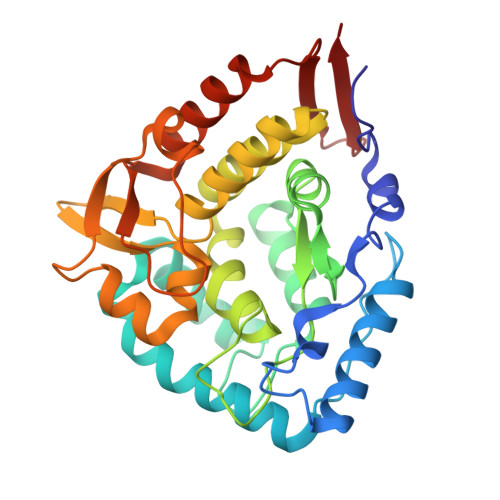
Crystal structure of phenylalanine hydroxylase (PAH) with Belinostat


Experimental Data Snapshot

  • Method: X-RAY DIFFRACTION
  • Resolution: 1.94 Å
  • R-Value Free: 
    0.203 (Depositor), 0.214 (DCC) 
  • R-Value Work: 
    0.166 (Depositor), 0.175 (DCC) 
  • R-Value Observed: 
    0.167 (Depositor) 

Starting Model: experimental
View more details

wwPDB Validation 3D Report Full Report

Validation slider image for 9T1O

Ligand Structure Quality Assessment 


This is version 1.0 of the entry. See complete history

Literature

Variant-dependent pharmacological rescue of phenylalanine hydroxylase supports a precision therapeutic strategy for phenylketonuria.

Conde-Gimenez, M.Salillas, S.Galiana-Cameo, M.Martinez-Olivan, J.E.Mahia, A.Ledesma, M.Galano-Frutos, J.J.Maity, R.Velazquez-Campoy, A.Diaz-de-Villegas, M.D.Hurtado-Guerrero, R.Sancho, J.

(2026) Biomed Pharmacother 199: 119371-119371

  • DOI: https://doi.org/10.1016/j.biopha.2026.119371
  • Primary Citation Related Structures: 
    9T1O

  • PubMed Abstract: 

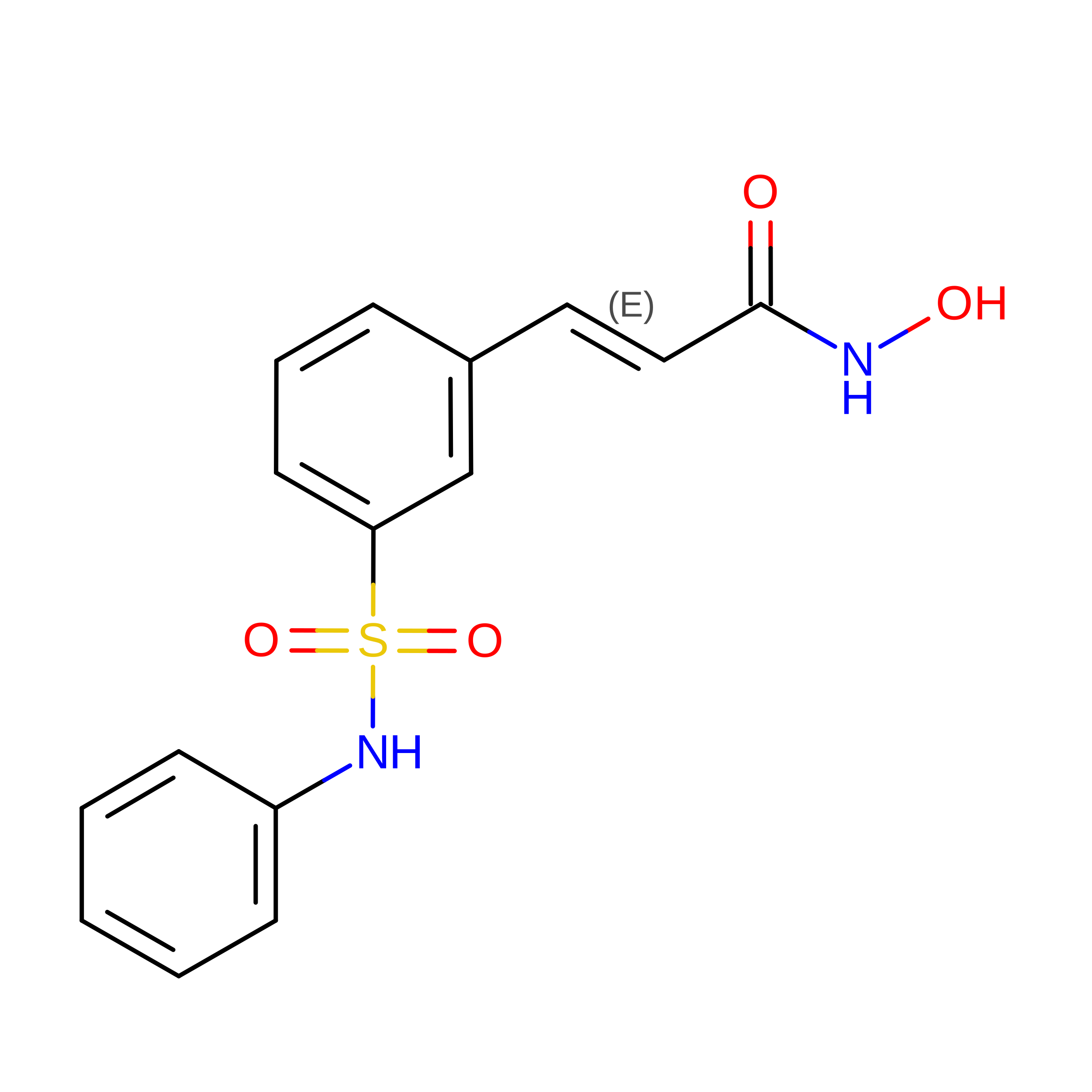
    Phenylketonuria (PKU) is an inherited metabolic disorder caused by pathogenic variants in phenylalanine hydroxylase (PAH), leading to toxic phenylalanine accumulation and severe neurological complications if untreated. Current pharmacological treatment relies on tetrahydrobiopterin (BH4), which benefits only a subset of patients, highlighting a major unmet need for alternative therapies. Here, we combined high-throughput screening, computational modelling, and drug repurposing to identify pharmacological chaperones capable of rescuing PAH function. We evaluated 26 structurally diverse small molecules in HEK293T cells expressing wild-type PAH or one of eight PKU-associated variants spanning phenotypes from mild to classical disease. Chaperoning efficacy was strongly variant-dependent, and for every variant tested at least one compound produced a greater activity increase than BH4 under identical assay conditions. Notably, belinostat, a clinically approved histone deacetylase inhibitor, emerged as the most effective compound for several clinically severe variants. Mechanistically, functional rescue consistently correlated with an increased population of tetrameric, catalytically competent PAH, as quantified by mass photometry. The crystal structure of the PAH-belinostat complex (PDB ID: 9T1O), together with structural models for all compounds, provide a framework for rational optimization. These results establish a preclinical proof-of-concept for genotype-guided pharmacological chaperone therapy in PKU and support the feasibility of personalized, variant-specific treatment strategies.


  • Organizational Affiliation
    • Departamento de Bioquímica y Biología Molecular y Celular, Facultad de Ciencias, Universidad de Zaragoza, Zaragoza 50009, Spain; Biocomputation and Complex Systems Physics Institute (BIFI)-GBsC-CSIC Joint Unit, Universidad de Zaragoza, Zaragoza 50018, Spain.

Macromolecule Content 

  • Total Structure Weight: 36.75 kDa 
  • Atom Count: 2,784 
  • Modeled Residue Count: 309 
  • Deposited Residue Count: 309 
  • Unique protein chains: 1

Macromolecules

Find similar proteins by:|  3D Structure
Entity ID: 1
MoleculeChains  Sequence LengthOrganismDetailsImage
Phenylalanine-4-hydroxylase309Homo sapiensMutation(s): 0 
Gene Names: PAH
EC: 1.14.16.1
UniProt & NIH Common Fund Data Resources
Find proteins for P00439 (Homo sapiens)
Explore P00439 
Go to UniProtKB:  P00439
PHAROS:  P00439
GTEx:  ENSG00000171759 
Entity Groups
Sequence Clusters30% Identity50% Identity70% Identity90% Identity95% Identity100% Identity
UniProt GroupP00439
Sequence Annotations
Expand
Reference Sequence

Small Molecules

Ligands 3 Unique
IDChains Name / Formula / InChI Key2D Diagram3D Interactions
5OG

Query on 5OG


Download:Ideal Coordinates CCD File
C [auth A]Belinostat
C15 H14 N2 O4 S
NCNRHFGMJRPRSK-MDZDMXLPSA-N
EDO

Query on EDO


Download:Ideal Coordinates CCD File
D [auth A]
E [auth A]
F [auth A]
G [auth A]
H [auth A]
D [auth A],
E [auth A],
F [auth A],
G [auth A],
H [auth A],
I [auth A],
J [auth A],
K [auth A],
L [auth A]
1,2-ETHANEDIOL
C2 H6 O2
LYCAIKOWRPUZTN-UHFFFAOYSA-N
FE

Query on FE


Download:Ideal Coordinates CCD File
B [auth A]FE (III) ION
Fe
VTLYFUHAOXGGBS-UHFFFAOYSA-N

Experimental Data & Validation

Experimental Data

  • Method: X-RAY DIFFRACTION
  • Resolution: 1.94 Å
  • R-Value Free:  0.203 (Depositor), 0.214 (DCC) 
  • R-Value Work:  0.166 (Depositor), 0.175 (DCC) 
  • R-Value Observed: 0.167 (Depositor) 
Space Group: C 2 2 21
Unit Cell:
Length ( Å )Angle ( ˚ )
a = 65.653α = 90
b = 108.103β = 90
c = 123.971γ = 90
Software Package:
Software NamePurpose
REFMACrefinement
XDSdata reduction
SCALAdata scaling
PHASERphasing

Structure Validation

View Full Validation Report


Ligand Structure Quality Assessment 


Entry History 

& Funding Information

Deposition Data


Funding OrganizationLocationGrant Number
Ministry of Economy and Competitiveness (MINECO)Spain--

Revision History  (Full details and data files)

  • Version 1.0: 2026-09-02
    Type: Initial release