A high diversity naive variable new antigen receptor, vNAR, phage library for rapid nanobody discovery across diverse antigens.
Kumar, V., Jangid, K., Santhosh, B., Dixit, R., Yadav, U., Verma, S., Rout, A., Surya, S., Das, M., Gupta, R., Saroj, A., Madhukalya, R., Gupta, M., Kalra, M., Iqbal, H., Kumar, D., Sinha, S., Tomar, S., Kumar, P., Kumar, R.(2025) J Biological Chem 302: 111083-111083
- PubMed: 41443416
- DOI: https://doi.org/10.1016/j.jbc.2025.111083
- Primary Citation Related Structures:
9UP9, 9UVH - PubMed Abstract:
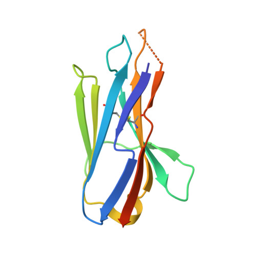
Conventional antibodies are among the most frequently used and effective biological tools explored for therapeutic and diagnostic applications. However, they face significant limitations when it comes to challenges that demand specialized attributes such as rapid tissue penetration, the ability to bind to concealed epitopes, and stability in non-physiological environments. In recent years, shark-derived immunoglobulin variable new antigen receptor (vNAR) has emerged as a promising alternative to overcome these limitations. In this study, we constructed a naïve vNAR phage display library from a white-spotted bamboo shark (Chiloscyllium plagiosum), with a library diversity size of ∼3 × 10 11 clones. Next generation sequencing analysis revealed the high diversity of the library, allowing it to encompass a broad range of classical functional vNAR types. To confirm the usability of the library for the successful isolation of positive clones, we screened the library against wide range of antigens (n=9;) from different origin that includes viral, cancer, autoimmune, toxins, parasite, algae and plant antigens. We achieved a hit rate of ∼100%, of potent binders with micro to nanomolar range affinity. The total number of unique binder's clones varied from 30%-100%, depending on the antigens and screening strategy. Furthermore, we provide an in-depth structural analysis by using X-ray crystallography of class IV vNARs from bamboo sharks, that remain underexplored. Our study represents a significant step forward in the field of single-domain antibody research and development.
- Department of Biosciences and Bioengineering, Indian Institute of Technology, Roorkee-247667, Uttarakhand, India.
Organizational Affiliation: