9V43 | pdb_00009v43

Cryo-EM structure of DICER/26S-UG complex in dicing state


Experimental Data Snapshot

  • Method: ELECTRON MICROSCOPY
  • Resolution: 3.34 Å
  • Aggregation State: PARTICLE 
  • Reconstruction Method: SINGLE PARTICLE 

Starting Model: experimental
View more details

wwPDB Validation 3D Report Full Report

Validation slider image for 9V43

This is version 1.1 of the entry. See complete history

Literature

DICER cleavage fidelity is governed by 5'-end binding pockets.

Ngo, M.K.Le, C.T.Nguyen, T.A.

(2026) Nature 

  • DOI: https://doi.org/10.1038/s41586-026-10211-5
  • Primary Citation Related Structures: 
    21CB, 21CN, 21CQ, 9V42, 9V43

  • PubMed Abstract: 

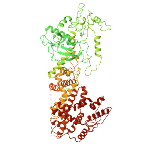
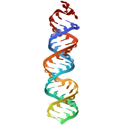
    RNA interference (RNAi) depends on DICER, an essential enzyme that processes RNA precursors into small regulatory RNAs. DICER cleaves RNA precursors according to the 5'-end counting rule, in which RNA length is measured from the 5'-end 1-3 . Previous work proposed a single 5'-end binding pocket that disfavours guanosine (5'-G), leading to cleavage inaccuracies 4 . Here we show that 5'-G promotes precise cleavage for many substrates. Using massively parallel dicing assays and cryo-electron microscopy, we identify a conserved guanosine-favoured (G-favoured) binding pocket in DICER, distinct from the previously described uridine-favoured (U-favoured) pocket. Together, these pockets influence the alignment between 21-nucleotide and 22-nucleotide cleavage registers, expanding the mechanism of small-RNA biogenesis in metazoan DICERs. We also find that conflicts between 5'-end binding and RNA-motif recognition can trigger RNA conformational adjustments that preserve accurate cleavage-site selection. In addition, conformational adjustments of the double-stranded RNA-binding domain (dsRBD) and PAZ domain help to align substrates with the catalytic centres for precise double-strand cleavage. These results show that the DICER cleavage mechanism integrates dual 5'-end binding pockets, RNA-motif influence and domain motions, advancing our understanding of microRNA biogenesis.


  • Organizational Affiliation
    • Division of Life Science, Hong Kong University of Science and Technology, Hong Kong, China.

Macromolecule Content 

  • Total Structure Weight: 238.61 kDa 
  • Atom Count: 6,889 
  • Modeled Residue Count: 766 
  • Deposited Residue Count: 1,973 
  • Unique protein chains: 1
  • Unique nucleic acid chains: 1

Macromolecules


Find similar proteins by:|  3D Structure
Entity ID: 2
MoleculeChains  Sequence LengthOrganismDetailsImage
Endoribonuclease Dicer1,909Homo sapiensMutation(s): 0 
Gene Names: DICER1DICERHERNAKIAA0928
EC: 3.1.26.3
UniProt & NIH Common Fund Data Resources
Find proteins for Q9UPY3 (Homo sapiens)
Explore Q9UPY3 
Go to UniProtKB:  Q9UPY3
PHAROS:  Q9UPY3
GTEx:  ENSG00000100697 
Entity Groups
Sequence Clusters30% Identity50% Identity70% Identity90% Identity95% Identity100% Identity
UniProt GroupQ9UPY3
Sequence Annotations
Expand
Reference Sequence
Find similar nucleic acids by:  (by identity cutoff) 
Entity ID: 1
MoleculeChains LengthOrganismImage
RNA (64-MER)64synthetic construct
Sequence Annotations
Expand
Reference Sequence

Experimental Data & Validation

Experimental Data

  • Method: ELECTRON MICROSCOPY
  • Resolution: 3.34 Å
  • Aggregation State: PARTICLE 
  • Reconstruction Method: SINGLE PARTICLE 
EM Software:
TaskSoftware PackageVersion
RECONSTRUCTIONcryoSPARC4.6.2
MODEL REFINEMENTPHENIX1.20.1_4487

Structure Validation

View Full Validation Report


Entry History 

& Funding Information

Deposition Data

  • Released Date: 2026-03-11 
  • Deposition Author(s): Ngo, M.K.

Funding OrganizationLocationGrant Number
Other governmentHong Kong16103023

Revision History  (Full details and data files)

  • Version 1.0: 2026-03-11
    Type: Initial release
  • Version 1.1: 2026-03-18
    Changes: Data collection, Database references