9W56 | pdb_00009w56

Structure of L-glutamate oxidase E617Q mutant in complex with L-glutamate


Experimental Data Snapshot

  • Method: X-RAY DIFFRACTION
  • Resolution: 2.38 Å
  • R-Value Free: 
    0.220 (Depositor), 0.222 (DCC) 
  • R-Value Work: 
    0.174 (Depositor), 0.177 (DCC) 
  • R-Value Observed: 
    0.177 (Depositor) 

Starting Model: experimental
View more details

wwPDB Validation   3D Report Full Report


Ligand Structure Quality Assessment 


This is version 1.0 of the entry. See complete history


Literature

Substrate recognition mechanisms of ʟ-glutamate oxidase from Streptomyces sp. and its conversion to ʟ-tyrosine oxidase.

Ueda, Y.Yano, Y.Nakayama, N.Takekawa, N.Inagaki, K.Imada, K.

(2026) Protein Sci 35: e70432-e70432

  • DOI: https://doi.org/10.1002/pro.70432
  • Primary Citation of Related Structures:  
    9W54, 9W55, 9W56, 9W57, 9W58

  • PubMed Abstract: 

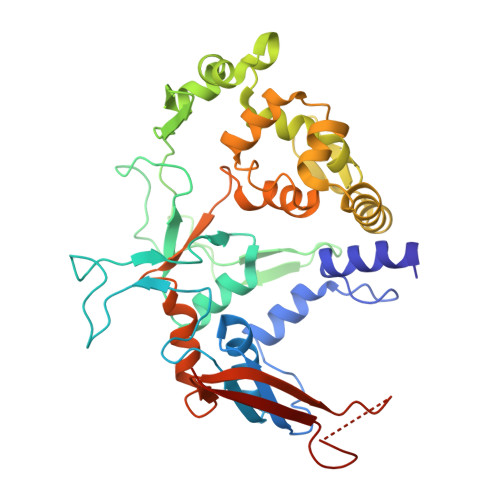
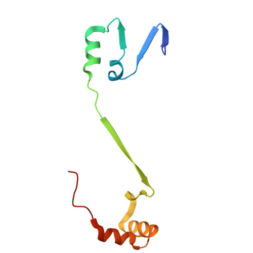
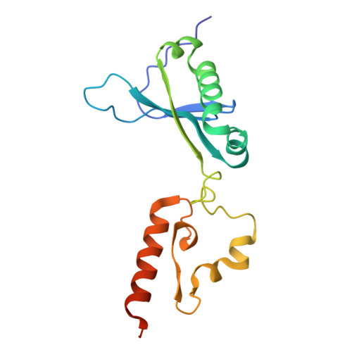
    ʟ-Amino acid oxidase (LAAO) is a flavoenzyme that catalyzes the oxidative deamination of ʟ-amino acids, producing α-keto acids, ammonia, and hydrogen peroxide. Among LAAOs, ʟ-glutamate oxidase (LGOX) from Streptomyces sp. X-119-6 exhibits exceptionally high substrate specificity for ʟ-glutamate. LGOX is expressed as a homodimeric precursor and undergoes proteolytic processing for maturation. Structural studies revealed that LGOX comprises an FAD-binding domain, a substrate-binding domain, and a helical domain. Conserved residues W653, R124, and Y562 that recognize the α-amino and α-carboxyl groups of the substrate exist in the putative active site. R305 was identified as a key determinant for side-chain recognition; its substitution with Glu conferred specific activity toward ʟ-arginine, effectively converting LGOX into an ʟ-arginine oxidase. However, the putative substrate binding pocket includes an acidic residue, E617, undesirable for acidic substrates. Therefore, the mechanism of high specificity for ʟ-glutamate remains unclear. To elucidate the molecular basis for the high substrate specificity of LGOX, we determined the structure of LGOX in complex with ʟ-glutamate. Structural and mutational analyses revealed that E617 plays a critical role in substrate binding by aligning the side chain of R305. The loop at the entrance of the tunnel to the substrate-binding site regulates the access of the substrate to the site. Furthermore, E617F and E617K variants acquired ʟ-tyrosine oxidase activity, providing insight into how specificity can be redirected. These findings clarify the substrate recognition mechanism of LGOX and underscore its potential as a robust scaffold for engineering novel amino acid oxidases with tailored specificities.


  • Organizational Affiliation
    • Department of Macromolecular Science, Graduate School of Science, The University of Osaka, Toyonaka, Osaka, Japan.

Macromolecules
Find similar proteins by:  (by identity cutoff)  |  3D Structure
Entity ID: 1
MoleculeChains Sequence LengthOrganismDetailsImage
L-glutamate oxidase precursor
A, D, G, J
376Streptomyces sp. X-119-6Mutation(s): 0 
Gene Names: lgoX
EC: 1.4.3.11
UniProt
Find proteins for Q8L3C7 (Streptomyces sp)
Explore Q8L3C7 
Go to UniProtKB:  Q8L3C7
Entity Groups  
Sequence Clusters30% Identity50% Identity70% Identity90% Identity95% Identity100% Identity
UniProt GroupQ8L3C7
Sequence Annotations
Expand
  • Reference Sequence
Find similar proteins by:  (by identity cutoff)  |  3D Structure
Entity ID: 2
MoleculeChains Sequence LengthOrganismDetailsImage
L-glutamate oxidase precursor
B, E, H, K
90Streptomyces sp. X-119-6Mutation(s): 0 
Gene Names: lgoX
EC: 1.4.3.11
UniProt
Find proteins for Q8L3C7 (Streptomyces sp)
Explore Q8L3C7 
Go to UniProtKB:  Q8L3C7
Entity Groups  
Sequence Clusters30% Identity50% Identity70% Identity90% Identity95% Identity100% Identity
UniProt GroupQ8L3C7
Sequence Annotations
Expand
  • Reference Sequence
Find similar proteins by:  (by identity cutoff)  |  3D Structure
Entity ID: 3
MoleculeChains Sequence LengthOrganismDetailsImage
L-glutamate oxidase precursor
C, F, I, L
163Streptomyces sp. X-119-6Mutation(s): 1 
Gene Names: lgoX
EC: 1.4.3.11
UniProt
Find proteins for Q8L3C7 (Streptomyces sp)
Explore Q8L3C7 
Go to UniProtKB:  Q8L3C7
Entity Groups  
Sequence Clusters30% Identity50% Identity70% Identity90% Identity95% Identity100% Identity
UniProt GroupQ8L3C7
Sequence Annotations
Expand
  • Reference Sequence
Small Molecules
Ligands 2 Unique
IDChains Name / Formula / InChI Key2D Diagram3D Interactions
FAD
Query on FAD

Download Ideal Coordinates CCD File 
M [auth A],
O [auth D],
Q [auth G],
S [auth J]
FLAVIN-ADENINE DINUCLEOTIDE
C27 H33 N9 O15 P2
VWWQXMAJTJZDQX-UYBVJOGSSA-N
GLU (Subject of Investigation/LOI)
Query on GLU

Download Ideal Coordinates CCD File 
N [auth A],
P [auth D],
R [auth G],
T [auth L]
GLUTAMIC ACID
C5 H9 N O4
WHUUTDBJXJRKMK-VKHMYHEASA-N
Experimental Data & Validation

Experimental Data

  • Method: X-RAY DIFFRACTION
  • Resolution: 2.38 Å
  • R-Value Free:  0.220 (Depositor), 0.222 (DCC) 
  • R-Value Work:  0.174 (Depositor), 0.177 (DCC) 
  • R-Value Observed: 0.177 (Depositor) 
Space Group: P 1 21 1
Unit Cell:
Length ( Å )Angle ( ˚ )
a = 85.957α = 90
b = 117.651β = 105.158
c = 147.953γ = 90
Software Package:
Software NamePurpose
PHENIXrefinement
MOSFLMdata reduction
Aimlessdata scaling
PHENIXphasing

Structure Validation

View Full Validation Report


Ligand Structure Quality Assessment 


Entry History & Funding Information

Deposition Data


Funding OrganizationLocationGrant Number
Japan Society for the Promotion of Science (JSPS)Japan21K05278

Revision History  (Full details and data files)

  • Version 1.0: 2026-01-07
    Type: Initial release