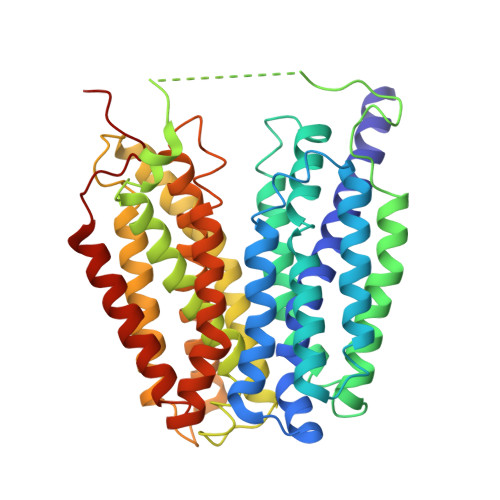
Structures of human glucose-6-phosphate transporter reveal reciprocal antiport mechanism driving glucose-6-phosphate and inorganic phosphate exchange.
Wang, Q., Guo, N., Du, Y., Liu, J., Ai, W., Yang, F., Zhu, Y., Zhao, Y., Wu, D., Liu, L., Yao, X., Gao, S.(2025) Nat Commun 16: 11441-11441
- PubMed: 41381426
- DOI: https://doi.org/10.1038/s41467-025-66386-4
- Primary Citation Related Structures:
9UGU, 9UGX, 9UGY, 9WS8, 9WT2, 9X8X, 9X95, 9X97 - PubMed Abstract:
Glucose-6-phosphate transporter 1 (G6PT1) is essential for systemic glucose homeostasis, and its deficiency causes glycogen storage disease type 1b (GSD1b). G6PT1 functions as a sugar-phosphate/inorganic phosphate (Pi) antiporter, orchestrating G6P transport into the endoplasmic reticulum lumen driven by a Pi gradient. Despite its physiological significance, the molecular mechanisms underlying substrate recognition and antiport activity remain poorly characterized. Here, we present cryo-electron microscopy structures of human G6PT1 in apo, Pi-bound, and GlcN6P-bound (a G6P analogue) states, all captured in cytosol-open conformations. Combined with molecular docking and functional assays, these structures elucidate the molecular basis for Pi and G6P recognition in G6PT1. Comparative analysis reveals that Pi binding triggers an interdomain salt bridge formation, resulting in a thicker luminal gate and a more compact central cavity for G6P binding. In addition to the monomer, we identify a dimeric assembly of G6PT1. Mutating key residues at the dimer interface impairs transport activity, suggesting a regulatory role for oligomerization. Our findings thus provide a mechanistic framework for understanding G6PT1 working mechanism and its pathological dysregulation in GSD1b.
- Department of Urology, Zhongnan Hospital of Wuhan University, State Key Laboratory of Metabolism and Regulation in Complex Organisms, School of Pharmaceutical Sciences, Wuhan University, Wuhan, China.
Organizational Affiliation: