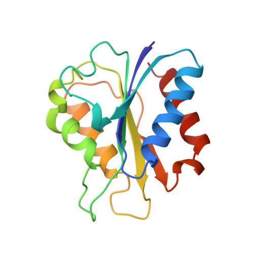
Crystallographic investigation of the role of aspartate 95 in the modulation of the redox potentials of Desulfovibrio vulgaris flavodoxin.
McCarthy, A.A., Walsh, M.A., Verma, C.S., O'Connell, D.P., Reinhold, M., Yalloway, G.N., D'Arcy, D., Higgins, T.M., Voordouw, G., Mayhew, S.G.(2002) Biochemistry 41: 10950-10962
- PubMed: 12206666
- DOI: https://doi.org/10.1021/bi020225h
- Primary Citation of Related Structures:
1AKQ, 1AKU, 1AKV, 1C7E, 1C7F - PubMed Abstract:
The side chain of aspartate 95 in flavodoxin from Desulfovibrio vulgaris provides the closest negative charge to N(1) of the bound FMN in the protein. Site-directed mutagenesis was used to substitute alanine, asparagine, or glutamate for this amino acid to assess the effect of this charge on the semiquinone/hydroquinone redox potential (E(1)) of the FMN cofactor. The D95A mutation shifts the E(1) redox potential positively by 16 mV, while a negative shift of 23 mV occurs in the oxidized/semiquinone midpoint redox potential (E(2)). The crystal structures of the oxidized and semiquinone forms of this mutant are similar to the corresponding states of the wild-type protein. In contrast to the wild-type protein, a further change in structure occurs in the D95A mutant in the hydroquinone form. The side chain of Y98 flips into an energetically more favorable edge-to-face interaction with the bound FMN. Analysis of the structural changes in the D95A mutant, taking into account electrostatic interactions at the FMN binding site, suggests that the pi-pi electrostatic repulsions have only a minor contribution to the very low E(1) redox potential of the FMN cofactor when bound to apoflavodoxin. Substitution of D95 with glutamate causes only a slight perturbation of the two one-electron redox potentials of the FMN cofactor. The structure of the D95E mutant reveals a large movement of the 60-loop (residues 60-64) away from the flavin in the oxidized structure. Reduction of this mutant to the hydroquinone causes the conformation of the 60-loop to revert back to that occurring in the structures of the wild-type protein. The crystal structures of the D95E mutant imply that electrostatic repulsion between a carboxylate on the side chain at position 95 and the phenol ring of Y98 prevents rotation of the Y98 side chain to a more energetically favorable conformation as occurs in the D95A mutant. Replacement of D95 with asparagine has no effect on E(2) but causes E(1) to change by 45 mV. The D95N mutant failed to crystallize. The K(d) values of the protein FMN complex in all three oxidation-reduction states differ from those of the wild-type complexes. Molecular modeling showed that the conformational energy of the protein changes with the redox state, in qualitative agreement with the observed changes in K(d), and allowed the electrostatic interactions between the FMN and the surrounding groups on the protein to be quantified.
- Department of Chemistry, National University of Ireland, Galway, Ireland.
Organizational Affiliation:


