1MIZ | pdb_00001miz

Crystal structure of an integrin beta3-talin chimera


Experimental Data Snapshot

  • Method: X-RAY DIFFRACTION
  • Resolution: 1.90 Å
  • R-Value Free: 
    0.246 (Depositor), 0.245 (DCC) 
  • R-Value Work: 
    0.204 (Depositor), 0.204 (DCC) 
  • R-Value Observed: 
    0.204 (Depositor) 

Starting Model: experimental
View more details

wwPDB Validation 3D Report Full Report

Validation slider image for 1MIZ

This is version 1.3 of the entry. See complete history

Literature

Structural determinants of integrin recognition by talin

Garcia-Alvarez, B.de Pereda, J.M.Calderwood, D.A.Ulmer, T.S.Critchley, D.Campbell, I.D.Ginsberg, M.H.Liddington, R.C.

(2003) Mol Cell 11: 49-58

  • DOI: https://doi.org/10.1016/s1097-2765(02)00823-7
  • Primary Citation Related Structures: 
    1MIX, 1MIZ, 1MK7, 1MK9

  • PubMed Abstract: 

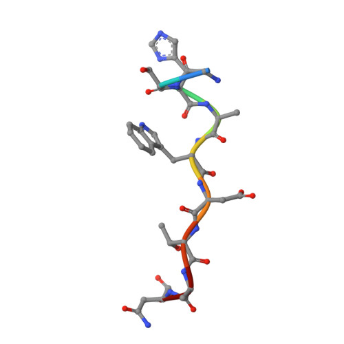
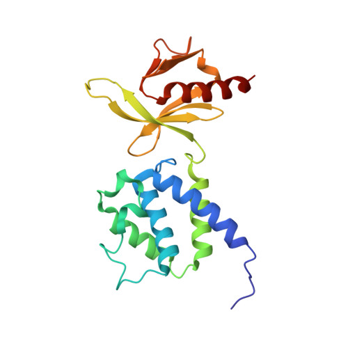
    The binding of cytoplasmic proteins, such as talin, to the cytoplasmic domains of integrin adhesion receptors mediates bidirectional signal transduction. Here we report the crystal structure of the principal integrin binding and activating fragment of talin, alone and in complex with fragments of the beta 3 integrin tail. The FERM (four point one, ezrin, radixin, and moesin) domain of talin engages integrins via a novel variant of the canonical phosphotyrosine binding (PTB) domain-NPxY ligand interaction that may be a prototype for FERM domain recognition of transmembrane receptors. In combination with NMR and mutational analysis, our studies reveal the critical interacting elements of both talin and the integrin beta 3 tail, providing structural paradigms for integrin linkage to the cell interior.


  • Organizational Affiliation
    • Program on Cell Adhesion, The Burnham Institute, 10901 North Torrey Pines Road, La Jolla, CA 92037, USA.

Macromolecule Content 

  • Total Structure Weight: 24.13 kDa 
  • Atom Count: 1,874 
  • Modeled Residue Count: 210 
  • Deposited Residue Count: 210 
  • Unique protein chains: 2

Macromolecules

Find similar proteins by:  Sequence   |   3D Structure  
Entity ID: 1
MoleculeChains  Sequence LengthOrganismDetailsImage
integrin beta39Homo sapiensMutation(s): 0 
UniProt & NIH Common Fund Data Resources
Find proteins for P05106 (Homo sapiens)
Explore P05106 
Go to UniProtKB:  P05106
PHAROS:  P05106
GTEx:  ENSG00000259207 
Entity Groups
UniProt GroupP05106
Sequence Annotations
Expand
Reference Sequence
Find similar proteins by:|  3D Structure
Entity ID: 2
MoleculeChains  Sequence LengthOrganismDetailsImage
TALIN201Gallus gallusMutation(s): 0 
UniProt
Find proteins for P54939 (Gallus gallus)
Explore P54939 
Go to UniProtKB:  P54939
Entity Groups
Sequence Clusters30% Identity50% Identity70% Identity90% Identity95% Identity100% Identity
UniProt GroupP54939
Sequence Annotations
Expand
Reference Sequence

Experimental Data & Validation

Experimental Data

  • Method: X-RAY DIFFRACTION
  • Resolution: 1.90 Å
  • R-Value Free:  0.246 (Depositor), 0.245 (DCC) 
  • R-Value Work:  0.204 (Depositor), 0.204 (DCC) 
  • R-Value Observed: 0.204 (Depositor) 
Space Group: P 1 21 1
Unit Cell:
Length ( Å )Angle ( ˚ )
a = 37.533α = 90
b = 55.198β = 97.58
c = 46.751γ = 90
Software Package:
Software NamePurpose
DENZOdata reduction
SCALEPACKdata scaling
MOLREPphasing
CNSrefinement
CCP4phasing

Structure Validation

View Full Validation Report


Entry History 

Deposition Data

Revision History  (Full details and data files)

  • Version 1.0: 2003-01-28
    Type: Initial release
  • Version 1.1: 2008-04-28
    Changes: Version format compliance
  • Version 1.2: 2011-07-13
    Changes: Version format compliance
  • Version 1.3: 2024-10-30
    Changes: Data collection, Database references, Derived calculations, Refinement description, Structure summary