1W2D

Human Inositol (1,4,5)-trisphosphate 3-kinase complexed with Mn2+/ADP/Ins(1,3,4,5)P4


Experimental Data Snapshot

  • Method: X-RAY DIFFRACTION
  • Resolution: 1.94 Å
  • R-Value Free: 
    0.267 (Depositor), 0.270 (DCC) 
  • R-Value Work: 
    0.226 (Depositor), 0.230 (DCC) 
  • R-Value Observed: 
    0.228 (Depositor) 

wwPDB Validation   3D Report Full Report


Ligand Structure Quality Assessment 

Created with Raphaël 2.3.0Worse 01 BetterLigand structure goodness of fit to experimental dataBest fitted 4IPClick on this verticalbar to view detailsBest fitted ADPClick on this verticalbar to view details

This is version 1.4 of the entry. See complete history


Literature

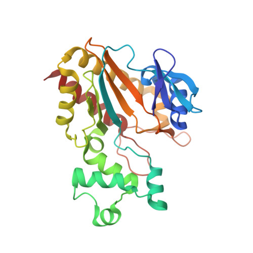
Structure of a Human Inositol 1,4,5-Trisphosphate 3-Kinase; Substrate Binding Reveals Why It is not a Phosphoinositide 3-Kinase

Gonzalez, B.Schell, M.J.Letcher, A.J.Veprintsev, D.B.Irvine, R.F.Williams, R.L.

(2004) Mol Cell 15: 689

  • DOI: https://doi.org/10.1016/j.molcel.2004.08.004
  • Primary Citation of Related Structures:  
    1W2C, 1W2D, 1W2F

  • PubMed Abstract: 

    Mammalian cells produce a variety of inositol phosphates (InsPs), including Ins(1,4,5)P3 that serves both as a second messenger and as a substrate for inositol polyphosphate kinases (IPKs), which further phosphorylate it. We report the structure of an IPK, the human Ins(1,4,5)P3 3-kinase-A, both free and in complexes with substrates and products. This enzyme catalyzes transfer of a phosphate from ATP to the 3-OH of Ins(1,4,5)P3, and its X-ray crystal structure provides a template for understanding a broad family of InsP kinases. The catalytic domain consists of three lobes. The N and C lobes bind ATP and resemble protein and lipid kinases, despite insignificant sequence similarity. The third lobe binds inositol phosphate and is a unique four-helix insertion in the C lobe. This lobe embraces all of the phosphates of Ins(1,4,5)P3 in a positively charged pocket, explaining the enzyme's substrate specificity and its inability to phosphorylate PtdIns(4,5)P2, the membrane-resident analog of Ins(1,4,5)P3.


  • Organizational Affiliation

    MRC Laboratory of Molecular Biology, Hills Road, Cambridge CB2 2QH, United Kingdom.


Macromolecules
Find similar proteins by:  (by identity cutoff)  |  3D Structure
Entity ID: 1
MoleculeChains Sequence LengthOrganismDetailsImage
INOSITOL-TRISPHOSPHATE 3-KINASE A
A, B
265Homo sapiensMutation(s): 0 
EC: 2.7.1.127
UniProt & NIH Common Fund Data Resources
Find proteins for P23677 (Homo sapiens)
Explore P23677 
Go to UniProtKB:  P23677
PHAROS:  P23677
GTEx:  ENSG00000137825 
Entity Groups  
Sequence Clusters30% Identity50% Identity70% Identity90% Identity95% Identity100% Identity
UniProt GroupP23677
Sequence Annotations
Expand
  • Reference Sequence
Small Molecules
Ligands 4 Unique
IDChains Name / Formula / InChI Key2D Diagram3D Interactions
4IP
Query on 4IP

Download Ideal Coordinates CCD File 
D [auth A],
I [auth B]
INOSITOL-(1,3,4,5)-TETRAKISPHOSPHATE
C6 H16 O18 P4
CIPFCGZLFXVXBG-CNWJWELYSA-N
ADP
Query on ADP

Download Ideal Coordinates CCD File 
C [auth A],
H [auth B]
ADENOSINE-5'-DIPHOSPHATE
C10 H15 N5 O10 P2
XTWYTFMLZFPYCI-KQYNXXCUSA-N
SO4
Query on SO4

Download Ideal Coordinates CCD File 
F [auth A],
G [auth A],
K [auth B]
SULFATE ION
O4 S
QAOWNCQODCNURD-UHFFFAOYSA-L
MN
Query on MN

Download Ideal Coordinates CCD File 
E [auth A],
J [auth B]
MANGANESE (II) ION
Mn
WAEMQWOKJMHJLA-UHFFFAOYSA-N
Binding Affinity Annotations 
IDSourceBinding Affinity
4IP BindingDB:  1W2D IC50: 2.78e+4 (nM) from 1 assay(s)
Experimental Data & Validation

Experimental Data

  • Method: X-RAY DIFFRACTION
  • Resolution: 1.94 Å
  • R-Value Free:  0.267 (Depositor), 0.270 (DCC) 
  • R-Value Work:  0.226 (Depositor), 0.230 (DCC) 
  • R-Value Observed: 0.228 (Depositor) 
Space Group: C 2 2 21
Unit Cell:
Length ( Å )Angle ( ˚ )
a = 72.054α = 90
b = 97.411β = 90
c = 190.844γ = 90
Software Package:
Software NamePurpose
REFMACrefinement
MOSFLMdata reduction
SCALAdata scaling

Structure Validation

View Full Validation Report


Ligand Structure Quality Assessment 

Created with Raphaël 2.3.0Worse 01 BetterLigand structure goodness of fit to experimental dataBest fitted 4IPClick on this verticalbar to view detailsBest fitted ADPClick on this verticalbar to view details

Entry History 

Deposition Data

Revision History  (Full details and data files)

  • Version 1.0: 2004-09-09
    Type: Initial release
  • Version 1.1: 2011-05-08
    Changes: Version format compliance
  • Version 1.2: 2011-07-13
    Changes: Version format compliance
  • Version 1.3: 2018-01-24
    Changes: Source and taxonomy
  • Version 1.4: 2024-05-08
    Changes: Data collection, Database references, Derived calculations, Other