1ZZU | pdb_00001zzu

Rat nNOS D597N/M336V double mutant with L-N(omega)-Nitroarginine-2,4-L-Diaminobutyric Amide Bound


Experimental Data Snapshot

  • Method: X-RAY DIFFRACTION
  • Resolution: 1.90 Å
  • R-Value Free: 
    0.228 (Depositor), 0.223 (DCC) 
  • R-Value Work: 
    0.202 (Depositor), 0.197 (DCC) 

wwPDB Validation 3D Report Full Report

Validation slider image for 1ZZU

Ligand Structure Quality Assessment 


This is version 1.5 of the entry. See complete history

Literature

Exploring the Binding Conformations of Bulkier Dipeptide Amide Inhibitors in Constitutive Nitric Oxide Synthases.

Li, H.Flinspach, M.L.Igarashi, J.Jamal, J.Yang, W.Litzinger, E.A.Huang, H.Erdal, E.P.Silverman, R.B.Poulos, T.L.

(2005) Biochemistry 44: 15222-15229

  • DOI: https://doi.org/10.1021/bi0513610
  • Primary Citation Related Structures: 
    1ZZQ, 1ZZR, 1ZZS, 1ZZT, 1ZZU

  • PubMed Abstract: 

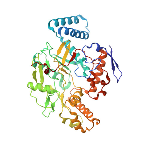
    A series of L-nitroarginine-based dipeptide inhibitors are highly selective for neuronal nitric oxide synthase (nNOS) over the endothelial isoform (eNOS). Crystal structures of these dipeptides bound to both isoforms revealed two different conformations, curled in nNOS and extended in eNOS, corresponding to higher and lower binding affinity to the two isoforms, respectively. In previous studies we found that the primary reason for selectivity is that Asp597 in nNOS, which is Asn368 in eNOS, provides greater electrostatic stabilization in the inhibitor complex. While this is the case for smaller dipeptide inhibitors, electrostatic stabilization may no longer be the sole determinant for isoform selectivity with bulkier dipeptide inhibitors. Another residue farther away from the active site, Met336 in nNOS (Val106 in eNOS), is in contact with bulkier dipeptide inhibitors. Double mutants were made to exchange the D597/M336 pair in nNOS with N368/V106 in eNOS. Here we report crystal structures and inhibition constants for bulkier dipeptide inhibitors bound to nNOS and eNOS that illustrate the important role played by residues near the entry to the active site in isoform selective inhibition.


  • Organizational Affiliation
    • Department of Molecular Biology and Biochemistry, University of California, Irvine, California 92697-3900, USA.

Macromolecule Content 

  • Total Structure Weight: 100.24 kDa 
  • Atom Count: 7,563 
  • Modeled Residue Count: 818 
  • Deposited Residue Count: 840 
  • Unique protein chains: 1

Macromolecules

Find similar proteins by:|  3D Structure
Entity ID: 1
MoleculeChains  Sequence LengthOrganismDetailsImage
Nitric-oxide synthase, brain
A, B
420Rattus norvegicusMutation(s): 2 
Gene Names: Nos1Bnos
EC: 1.14.13.39
UniProt
Find proteins for P29476 (Rattus norvegicus)
Explore P29476 
Go to UniProtKB:  P29476
Entity Groups
Sequence Clusters30% Identity50% Identity70% Identity90% Identity95% Identity100% Identity
UniProt GroupP29476
Sequence Annotations
Expand
Reference Sequence

Small Molecules

Ligands 7 Unique
IDChains Name / Formula / InChI Key2D Diagram3D Interactions
HEM

Query on HEM


Download:Ideal Coordinates CCD File
F [auth A],
L [auth B]
PROTOPORPHYRIN IX CONTAINING FE
C34 H32 Fe N4 O4
KABFMIBPWCXCRK-RGGAHWMASA-L
DP1

Query on DP1


Download:Ideal Coordinates CCD File
H [auth A],
N [auth B]
L-N(OMEGA)-NITROARGININE-2,4-L-DIAMINOBUTYRIC AMIDE
C10 H22 N8 O4
KUZKVXUOMSVPOA-BQBZGAKWSA-N
H4B

Query on H4B


Download:Ideal Coordinates CCD File
G [auth A],
M [auth B]
5,6,7,8-TETRAHYDROBIOPTERIN
C9 H15 N5 O3
FNKQXYHWGSIFBK-RPDRRWSUSA-N
MTL

Query on MTL


Download:Ideal Coordinates CCD File
E [auth A],
K [auth B]
D-MANNITOL
C6 H14 O6
FBPFZTCFMRRESA-KVTDHHQDSA-N
GOL

Query on GOL


Download:Ideal Coordinates CCD File
I [auth A],
O [auth B]
GLYCEROL
C3 H8 O3
PEDCQBHIVMGVHV-UHFFFAOYSA-N
ZN

Query on ZN


Download:Ideal Coordinates CCD File
D [auth A]ZINC ION
Zn
PTFCDOFLOPIGGS-UHFFFAOYSA-N
ACT

Query on ACT


Download:Ideal Coordinates CCD File
C [auth A],
J [auth B]
ACETATE ION
C2 H3 O2
QTBSBXVTEAMEQO-UHFFFAOYSA-M

Experimental Data & Validation

Experimental Data

  • Method: X-RAY DIFFRACTION
  • Resolution: 1.90 Å
  • R-Value Free:  0.228 (Depositor), 0.223 (DCC) 
  • R-Value Work:  0.202 (Depositor), 0.197 (DCC) 
Space Group: P 21 21 21
Unit Cell:
Length ( Å )Angle ( ˚ )
a = 52.36α = 90
b = 111.13β = 90
c = 165.16γ = 90
Software Package:
Software NamePurpose
DENZOdata reduction
SCALEPACKdata scaling
CNSrefinement
PDB_EXTRACTdata extraction
HKL-2000data reduction
CNSphasing

Structure Validation

View Full Validation Report


Ligand Structure Quality Assessment 


Entry History 

Deposition Data

Revision History  (Full details and data files)

  • Version 1.0: 2005-12-06
    Type: Initial release
  • Version 1.1: 2008-04-30
    Changes: Version format compliance
  • Version 1.2: 2011-07-13
    Changes: Non-polymer description, Version format compliance
  • Version 1.3: 2017-10-11
    Changes: Refinement description
  • Version 1.4: 2021-10-20
    Changes: Database references, Derived calculations
  • Version 1.5: 2024-02-14
    Changes: Data collection