2B0M | pdb_00002b0m

Human dihydroorotate dehydrogenase bound to a novel inhibitor


Experimental Data Snapshot

  • Method: X-RAY DIFFRACTION
  • Resolution: 2.00 Å
  • R-Value Free: 
    0.249 (Depositor), 0.250 (DCC) 
  • R-Value Work: 
    0.218 (Depositor), 0.219 (DCC) 

Starting Model: experimental
View more details

wwPDB Validation 3D Report Full Report

Validation slider image for 2B0M

Ligand Structure Quality Assessment 


This is version 1.3 of the entry. See complete history

Literature

Brequinar derivatives and species-specific drug design for dihydroorotate dehydrogenase.

Hurt, D.E.Sutton, A.E.Clardy, J.

(2006) Bioorg Med Chem Lett 16: 1610-1615

  • DOI: https://doi.org/10.1016/j.bmcl.2005.12.029
  • Primary Citation Related Structures: 
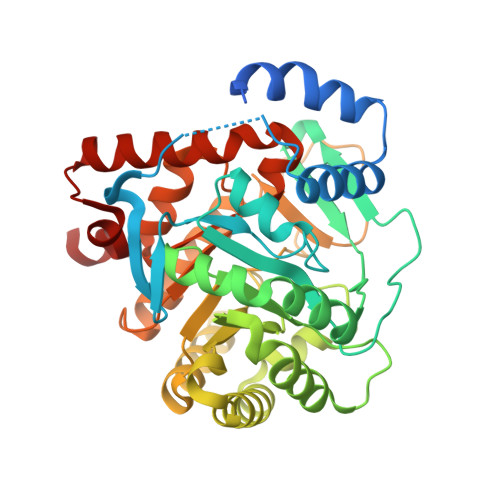
    2B0M

  • PubMed Abstract: 

    Therapeutic agents brequinar sodium and leflunomide (Arava) work by binding in a hydrophobic tunnel formed by a highly variable N-terminus of family 2 dihydroorotate dehydrogenase (DHODH). The X-ray crystallographic structure of an analog of brequinar bound to human DHODH was determined. In silico screening of a library of compounds suggested another subset of brequinar analogs that do not inhibit human DHODH as potentially effective inhibitors of Plasmodium falciparum DHODH.


  • Organizational Affiliation
    • Department of Chemistry and Chemical Biology, Cornell University, Ithaca, NY 14850, USA.

Macromolecule Content 

  • Total Structure Weight: 43.94 kDa 
  • Atom Count: 2,964 
  • Modeled Residue Count: 361 
  • Deposited Residue Count: 393 
  • Unique protein chains: 1

Macromolecules

Find similar proteins by:|  3D Structure
Entity ID: 1
MoleculeChains  Sequence LengthOrganismDetailsImage
Dihydroorotate dehydrogenase, mitochondrial393Homo sapiensMutation(s): 0 
Gene Names: DHODH
EC: 1.3.3.1 (PDB Primary Data), 1.3.5.2 (UniProt)
UniProt & NIH Common Fund Data Resources
Find proteins for Q02127 (Homo sapiens)
Explore Q02127 
Go to UniProtKB:  Q02127
PHAROS:  Q02127
GTEx:  ENSG00000102967 
Entity Groups
Sequence Clusters30% Identity50% Identity70% Identity90% Identity95% Identity100% Identity
UniProt GroupQ02127
Sequence Annotations
Expand
Reference Sequence

Experimental Data & Validation

Experimental Data

  • Method: X-RAY DIFFRACTION
  • Resolution: 2.00 Å
  • R-Value Free:  0.249 (Depositor), 0.250 (DCC) 
  • R-Value Work:  0.218 (Depositor), 0.219 (DCC) 
Space Group: P 32 2 1
Unit Cell:
Length ( Å )Angle ( ˚ )
a = 90.398α = 90
b = 90.398β = 90
c = 123.332γ = 120
Software Package:
Software NamePurpose
HKL-2000data collection
SCALEPACKdata scaling
MOLREPphasing
CNSrefinement
HKL-2000data reduction

Structure Validation

View Full Validation Report


Ligand Structure Quality Assessment 


Entry History 

Deposition Data

Revision History  (Full details and data files)

  • Version 1.0: 2005-09-27
    Type: Initial release
  • Version 1.1: 2008-05-01
    Changes: Version format compliance
  • Version 1.2: 2011-07-13
    Changes: Version format compliance
  • Version 1.3: 2023-08-23
    Changes: Data collection, Database references, Derived calculations, Refinement description