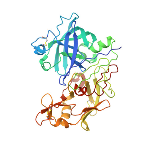
2V12

Crystal Structure of Renin with Inhibitor 8


Experimental Data Snapshot

  • Method: X-RAY DIFFRACTION
  • Resolution: 3.20 Å
  • R-Value Work: 0.236 
  • R-Value Observed: 0.236 

wwPDB Validation   3D Report Full Report


This is version 1.6 of the entry. See complete history


Literature

Structure-Based Drug Design: The Discovery of Novel Nonpeptide Orally Active Inhibitors of Human Renin

Rahuel, J.Rasetti, V.Maibaum, J.Rueger, H.Goschke, R.Cohen, N.C.Stutz, S.Cumin, F.Fuhrer, W.Wood, J.M.Grutter, M.G.

(2000) Chem Biol 7: 493

  • DOI: https://doi.org/10.1016/s1074-5521(00)00134-4
  • Primary Citation of Related Structures:  
    2V0Z, 2V10, 2V11, 2V12, 2V13, 2V16

  • PubMed Abstract: 

    The aspartic proteinase renin plays an important physiological role in the regulation of blood pressure. It catalyses the first step in the conversion of angiotensinogen to the hormone angiotensin II. In the past, potent peptide inhibitors of renin have been developed, but none of these compounds has made it to the end of clinical trials. Our primary aim was to develop novel nonpeptide inhibitors. Based on the available structural information concerning renin-substrate interactions, we synthesized inhibitors in which the peptide portion was replaced by lipophilic moieties that interact with the large hydrophobic S1/S3-binding pocket in renin. Crystal structure analysis of renin-inhibitor complexes combined with computational methods were employed in the medicinal-chemistry optimisation process. Structure analysis revealed that the newly designed inhibitors bind as predicted to the S1/S3 pocket. In addition, however, these compounds interact with a hitherto unrecognised large, distinct, sub-pocket of the enzyme that extends from the S3-binding site towards the hydrophobic core of the enzyme. Binding to this S3(sp) sub-pocket was essential for high binding affinity. This unprecedented binding mode guided the drug-design process in which the mostly hydrophobic interactions within subsite S3(sp) were optimised. Our design approach led to compounds with high in vitro affinity and specificity for renin, favourable bioavailability and excellent oral efficacy in lowering blood pressure in primates. These renin inhibitors are therefore potential therapeutic agents for the treatment of hypertension and related cardiovascular diseases.


  • Organizational Affiliation

    Core Technology Area, Novartis Pharma AG, Metabolic and Cardiovascular Diseases, Basle, Switzerland.


Macromolecules
Find similar proteins by:  (by identity cutoff)  |  3D Structure
Entity ID: 1
MoleculeChains Sequence LengthOrganismDetailsImage
RENINA [auth C],
B [auth O]
340Homo sapiensMutation(s): 0 
EC: 3.4.23.15
UniProt & NIH Common Fund Data Resources
Find proteins for P00797 (Homo sapiens)
Explore P00797 
Go to UniProtKB:  P00797
PHAROS:  P00797
GTEx:  ENSG00000143839 
Entity Groups  
Sequence Clusters30% Identity50% Identity70% Identity90% Identity95% Identity100% Identity
UniProt GroupP00797
Sequence Annotations
Expand
  • Reference Sequence
Small Molecules
Ligands 1 Unique
IDChains Name / Formula / InChI Key2D Diagram3D Interactions
C39
Query on C39

Download Ideal Coordinates CCD File 
C,
D [auth O]
N-[(2S,4S,5S,7R)-4-AMINO-8-(BUTYLAMINO)-5-HYDROXY-7-METHYL-2-(1-METHYLETHYL)-8-OXOOCTYL]-2-(3-METHOXYPROPOXY)BENZAMIDE
C27 H47 N3 O5
JYPXLSXKONMTFW-HTDNTCHWSA-N
Binding Affinity Annotations 
IDSourceBinding Affinity
C39 PDBBind:  2V12 IC50: 0.9 (nM) from 1 assay(s)
BindingDB:  2V12 IC50: 0.9 (nM) from 1 assay(s)
Experimental Data & Validation

Experimental Data

  • Method: X-RAY DIFFRACTION
  • Resolution: 3.20 Å
  • R-Value Work: 0.236 
  • R-Value Observed: 0.236 
  • Space Group: P 21 3
Unit Cell:
Length ( Å )Angle ( ˚ )
a = 142.9α = 90
b = 142.9β = 90
c = 142.9γ = 90
Software Package:
Software NamePurpose
X-PLORrefinement
MADNESSdata reduction

Structure Validation

View Full Validation Report


Entry History 

Deposition Data

Revision History  (Full details and data files)

  • Version 1.0: 2007-07-03
    Type: Initial release
  • Version 1.1: 2011-05-08
    Changes: Version format compliance
  • Version 1.2: 2011-07-13
    Changes: Version format compliance
  • Version 1.3: 2017-06-28
    Changes: Data collection
  • Version 1.4: 2017-07-12
    Changes: Refinement description
  • Version 1.5: 2019-04-03
    Changes: Data collection, Source and taxonomy
  • Version 1.6: 2024-11-13
    Changes: Data collection, Database references, Derived calculations, Structure summary