3A37

Structural insight into the membrane insertion of tail-anchored proteins by Get3


Experimental Data Snapshot

  • Method: X-RAY DIFFRACTION
  • Resolution: 3.00 Å
  • R-Value Free: 
    0.282 (Depositor), 0.270 (DCC) 
  • R-Value Work: 
    0.253 (Depositor), 0.240 (DCC) 
  • R-Value Observed: 
    0.253 (Depositor) 

wwPDB Validation   3D Report Full Report


Ligand Structure Quality Assessment 

Created with Raphaël 2.3.0Worse 01 BetterLigand structure goodness of fit to experimental dataBest fitted ADPClick on this verticalbar to view details

This is version 1.3 of the entry. See complete history


Literature

Structural insight into the membrane insertion of tail-anchored proteins by Get3

Yamagata, A.Mimura, H.Sato, Y.Yamashita, M.Yoshikawa, A.Fukai, S.

(2010) Genes Cells 15: 29-41

  • DOI: https://doi.org/10.1111/j.1365-2443.2009.01362.x
  • Primary Citation of Related Structures:  
    3A36, 3A37

  • PubMed Abstract: 

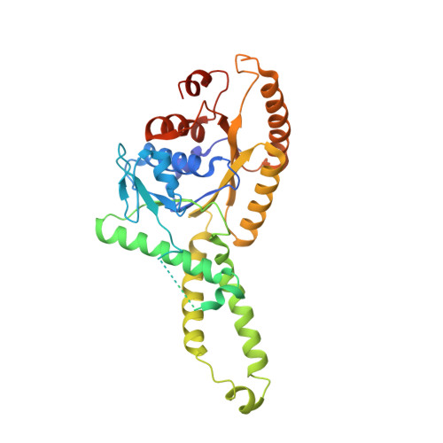
    Tail anchored (TA) proteins, which are important for numerous cellular processes, are defined by a single transmembrane domain (TMD) near the C-terminus. The membrane insertion of TA proteins is mediated by the highly conserved ATPase Get3. Here we report the crystal structures of Get3 in ADP-bound and nucleotide-free forms at 3.0 A and 2.8 A resolutions, respectively. Get3 consists of a nucleotide binding domain and a helical domain. Both structures exhibit a Zn(2+)-mediated homodimer in a head-to-head orientation, representing an open dimer conformation. Our cross-link experiments indicated the closed dimer-stimulating ATP hydrolysis, which might be coupled with TA-protein release. Further, our coexpression-based binding assays using a model TA protein Sec22p revealed the direct interaction between the helical domain of Get3 and the Sec22p TMD. This interaction is independent of ATP and dimer formation. Finally, we propose a structural mechanism that links ATP hydrolysis with the TA-protein insertion mediated by the conserved DTAPTGH motif.


  • Organizational Affiliation

    Structural Biology Laboratory, Life Science Division, Synchrotron Radiation Research Organization and Institute of Molecular and Cellular Biosciences, The University of Tokyo, Tokyo 113-0032, Japan.


Macromolecules
Find similar proteins by:  (by identity cutoff)  |  3D Structure
Entity ID: 1
MoleculeChains Sequence LengthOrganismDetailsImage
ATPase GET3
A, B
362Saccharomyces cerevisiaeMutation(s): 0 
Gene Names: 851458
EC: 3.6.3.16 (PDB Primary Data), 3.6 (UniProt)
UniProt
Find proteins for Q12154 (Saccharomyces cerevisiae (strain ATCC 204508 / S288c))
Explore Q12154 
Go to UniProtKB:  Q12154
Entity Groups  
Sequence Clusters30% Identity50% Identity70% Identity90% Identity95% Identity100% Identity
UniProt GroupQ12154
Sequence Annotations
Expand
  • Reference Sequence
Experimental Data & Validation

Experimental Data

  • Method: X-RAY DIFFRACTION
  • Resolution: 3.00 Å
  • R-Value Free:  0.282 (Depositor), 0.270 (DCC) 
  • R-Value Work:  0.253 (Depositor), 0.240 (DCC) 
  • R-Value Observed: 0.253 (Depositor) 
Space Group: P 21 21 2
Unit Cell:
Length ( Å )Angle ( ˚ )
a = 115.229α = 90
b = 222.554β = 90
c = 49.291γ = 90
Software Package:
Software NamePurpose
CNSrefinement
HKL-2000data collection
HKL-2000data reduction
HKL-2000data scaling
CNSphasing

Structure Validation

View Full Validation Report


Ligand Structure Quality Assessment 

Created with Raphaël 2.3.0Worse 01 BetterLigand structure goodness of fit to experimental dataBest fitted ADPClick on this verticalbar to view details

Entry History 

Deposition Data

Revision History  (Full details and data files)

  • Version 1.0: 2010-01-12
    Type: Initial release
  • Version 1.1: 2011-07-13
    Changes: Version format compliance
  • Version 1.2: 2018-02-14
    Changes: Experimental preparation
  • Version 1.3: 2024-03-13
    Changes: Data collection, Database references, Derived calculations