3EG6

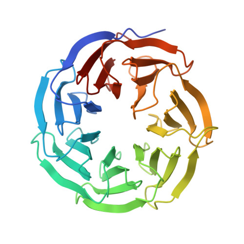
Structure of WDR5 bound to MLL1 peptide


Experimental Data Snapshot

  • Method: X-RAY DIFFRACTION
  • Resolution: 1.72 Å
  • R-Value Free: 0.240 
  • R-Value Work: 0.203 
  • R-Value Observed: 0.203 

Starting Model: experimental
View more details

wwPDB Validation   3D Report Full Report


This is version 1.4 of the entry. See complete history


Literature

Structure of WDR5 bound to mixed lineage leukemia protein-1 peptide.

Patel, A.Dharmarajan, V.Cosgrove, M.S.

(2008) J Biol Chem 283: 32158-32161

  • DOI: https://doi.org/10.1074/jbc.C800164200
  • Primary Citation of Related Structures:  
    3EG6

  • PubMed Abstract: 

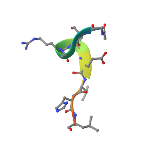
    The mixed lineage leukemia protein-1 (MLL1) catalyzes histone H3 lysine 4 methylation and is regulated by interaction with WDR5 (WD-repeat protein-5), RbBP5 (retinoblastoma-binding protein-5), and the Ash2L (absent, small, homeotic discs-2-like) oncoprotein. In the accompanying investigation, we describe the identification of a conserved arginine containing motif, called the "Win" or WDR5 interaction motif, that is essential for the assembly and H3K4 dimethylation activity of the MLL1 core complex. Here we present a 1.7-A crystal structure of WDR5 bound to a peptide derived from the MLL1 Win motif. Our results show that Arg-3765 of MLL1 is bound in the same arginine binding pocket on WDR5 that was previously suggested to bind histone H3. Thermodynamic binding experiments show that the MLL1 Win peptide is preferentially recognized by WDR5. These results are consistent with a model in which WDR5 recognizes Arg-3765 of MLL1, which is essential for the assembly and enzymatic activity of the MLL1 core complex.


  • Organizational Affiliation

    Department of Biology, Syracuse University, Syracuse, New York 13244, USA.


Macromolecules
Find similar proteins by:  (by identity cutoff)  |  3D Structure
Entity ID: 1
MoleculeChains Sequence LengthOrganismDetailsImage
WD repeat-containing protein 5312Homo sapiensMutation(s): 0 
Gene Names: WDR5BIG3
UniProt & NIH Common Fund Data Resources
Find proteins for P61964 (Homo sapiens)
Explore P61964 
Go to UniProtKB:  P61964
PHAROS:  P61964
GTEx:  ENSG00000196363 
Entity Groups  
Sequence Clusters30% Identity50% Identity70% Identity90% Identity95% Identity100% Identity
UniProt GroupP61964
Sequence Annotations
Expand
  • Reference Sequence

Find similar proteins by:  Sequence   |   3D Structure  

Entity ID: 2
MoleculeChains Sequence LengthOrganismDetailsImage
MLL-1 peptideB [auth C]13N/AMutation(s): 0 
EC: 2.1.1.364 (UniProt), 2.1.1 (UniProt)
UniProt & NIH Common Fund Data Resources
Find proteins for Q03164 (Homo sapiens)
Explore Q03164 
Go to UniProtKB:  Q03164
PHAROS:  Q03164
GTEx:  ENSG00000118058 
Entity Groups  
Sequence Clusters30% Identity50% Identity70% Identity90% Identity95% Identity100% Identity
UniProt GroupQ03164
Sequence Annotations
Expand
  • Reference Sequence
Experimental Data & Validation

Experimental Data

Unit Cell:
Length ( Å )Angle ( ˚ )
a = 78.254α = 90
b = 98.384β = 90
c = 80.075γ = 90
Software Package:
Software NamePurpose
DENZOdata reduction
SCALEPACKdata scaling
MOLREPphasing
CNSrefinement
PDB_EXTRACTdata extraction
HKL-2000data reduction

Structure Validation

View Full Validation Report


Entry History 

Deposition Data

Revision History  (Full details and data files)

  • Version 1.0: 2008-09-30
    Type: Initial release
  • Version 1.1: 2011-07-13
    Changes: Version format compliance
  • Version 1.2: 2017-10-25
    Changes: Refinement description
  • Version 1.3: 2023-08-30
    Changes: Data collection, Database references, Derived calculations, Refinement description
  • Version 1.4: 2024-10-16
    Changes: Structure summary