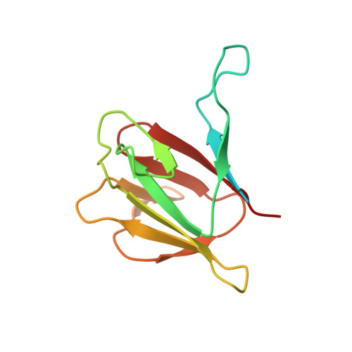
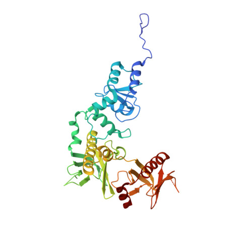
3FM8 | pdb_00003fm8

Crystal structure of full length centaurin alpha-1 bound with the FHA domain of KIF13B (CAPRI target)


Experimental Data Snapshot

  • Method: X-RAY DIFFRACTION
  • Resolution: 2.30 Å
  • R-Value Free: 
    0.272 (Depositor), 0.271 (DCC) 
  • R-Value Work: 
    0.231 (Depositor), 0.233 (DCC) 
  • R-Value Observed: 
    0.232 (Depositor) 

Starting Model: other
View more details

wwPDB Validation 3D Report Full Report

Validation slider image for 3FM8

This is version 1.5 of the entry. See complete history. 

Literature

Phosphorylation-independent dual-site binding of the FHA domain of KIF13 mediates phosphoinositide transport via centaurin alpha1.

Tong, Y., Tempel, W., Wang, H., Yamada, K., Shen, L., Senisterra, G.A., MacKenzie, F., Chishti, A.H., Park, H.W.

(2010) Proc Natl Acad Sci U S A 107: 20346-20351

  • DOI: https://doi.org/10.1073/pnas.1009008107
  • Primary Citation Related Structures: 
    3FEH, 3FM8, 3LJU

  • PubMed Abstract: 

    Phosphatidylinositol 3,4,5-triphosphate (PIP3) plays a key role in neuronal polarization and axon formation. PIP3-containing vesicles are transported to axon tips by the kinesin KIF13B via an adaptor protein, centaurin α1 (CENTA1). KIF13B interacts with CENTA1 through its forkhead-associated (FHA) domain. We solved the crystal structures of CENTA1 in ligand-free, KIF13B-FHA domain-bound, and PIP3 head group (IP4)-bound conformations, and the CENTA1/KIF13B-FHA/IP4 ternary complex. The first pleckstrin homology (PH) domain of CENTA1 specifically binds to PIP3, while the second binds to both PIP3 and phosphatidylinositol 3,4-biphosphate (PI(3,4)P(2)). The FHA domain of KIF13B interacts with the PH1 domain of one CENTA1 molecule and the ArfGAP domain of a second CENTA1 molecule in a threonine phosphorylation-independent fashion. We propose that full-length KIF13B and CENTA1 form heterotetramers that can bind four phosphoinositide molecules in the vesicle and transport it along the microtubule.


  • Organizational Affiliation: 
    • Structural Genomics Consortium, University of Toronto, Toronto, ON M5G 1L7, Canada.

Macromolecule Content 

  • Total Structure Weight: 119.75 kDa 
  • Atom Count: 7,335 
  • Modeled Residue Count: 910 
  • Deposited Residue Count: 1,032 
  • Unique protein chains: 2

Macromolecules

Find similar proteins by:|  3D Structure
Entity ID: 1
MoleculeChains  Sequence LengthOrganismDetailsImage
Kinesin-like protein KIF13B
A, B
124Homo sapiensMutation(s): 0 
Gene Names: KIF13B, GAKIN, KIAA0639
UniProt & NIH Common Fund Data Resources
Find proteins for Q9NQT8 (Homo sapiens)
Explore Q9NQT8 
Go to UniProtKB:  Q9NQT8
PHAROS:  Q9NQT8
GTEx:  ENSG00000197892 
Entity Groups
Sequence Clusters30% Identity50% Identity70% Identity90% Identity95% Identity100% Identity
UniProt GroupQ9NQT8
Sequence Annotations
Expand
Reference Sequence
Find similar proteins by:|  3D Structure
Entity ID: 2
MoleculeChains  Sequence LengthOrganismDetailsImage
Centaurin-alpha-1
C, D
392Homo sapiensMutation(s): 0 
Gene Names: CENTA1
UniProt & NIH Common Fund Data Resources
Find proteins for O75689 (Homo sapiens)
Explore O75689 
Go to UniProtKB:  O75689
PHAROS:  O75689
GTEx:  ENSG00000105963 
Entity Groups
Sequence Clusters30% Identity50% Identity70% Identity90% Identity95% Identity100% Identity
UniProt GroupO75689
Sequence Annotations
Expand
Reference Sequence

Small Molecules

Ligands 3 Unique
IDChains Name / Formula / InChI Key2D Diagram3D Interactions
SO4

Query on SO4


Download:Ideal Coordinates CCD File
J [auth C],
R [auth D],
S [auth D]
SULFATE ION
O4 S
QAOWNCQODCNURD-UHFFFAOYSA-L
ZN

Query on ZN


Download:Ideal Coordinates CCD File
I [auth C],
Q [auth D]
ZINC ION
Zn
PTFCDOFLOPIGGS-UHFFFAOYSA-N
UNX

Query on UNX


Download:Ideal Coordinates CCD File
AA [auth D]
BA [auth D]
CA [auth D]
DA [auth D]
E [auth A]
AA [auth D],
BA [auth D],
CA [auth D],
DA [auth D],
E [auth A],
EA [auth D],
F [auth A],
G [auth A],
H [auth B],
K [auth C],
L [auth C],
M [auth C],
N [auth C],
O [auth C],
P [auth C],
T [auth D],
U [auth D],
V [auth D],
W [auth D],
X [auth D],
Y [auth D],
Z [auth D]
UNKNOWN ATOM OR ION
X

Experimental Data & Validation

Experimental Data

  • Method: X-RAY DIFFRACTION
  • Resolution: 2.30 Å
  • R-Value Free:  0.272 (Depositor), 0.271 (DCC) 
  • R-Value Work:  0.231 (Depositor), 0.233 (DCC) 
  • R-Value Observed: 0.232 (Depositor) 
Space Group: P 41 21 2
Unit Cell:
Length ( Å )Angle ( ˚ )
a = 115.632α = 90
b = 115.632β = 90
c = 189.291γ = 90
Software Package:
Software NamePurpose
DENZOdata reduction
SCALEPACKdata scaling
MOLREPphasing
REFMACrefinement
PDB_EXTRACTdata extraction
HKL-2000data scaling

Structure Validation

View Full Validation Report


Entry History 

Revision History  (Full details and data files)

  • Version 1.0: 2009-08-25
    Type: Initial release
  • Version 1.1: 2011-07-13
    Changes: Version format compliance
  • Version 1.2: 2013-09-25
    Changes: Derived calculations
  • Version 1.3: 2017-11-01
    Changes: Refinement description
  • Version 1.4: 2024-02-21
    Changes: Data collection, Database references, Derived calculations
  • Version 1.5: 2024-04-03
    Changes: Refinement description