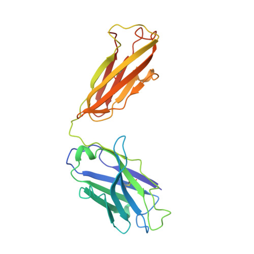
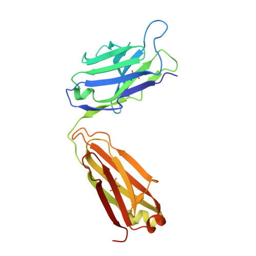
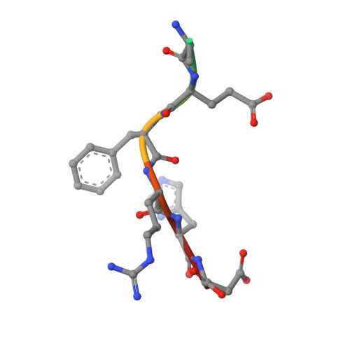
3IFP

X-ray structure of amyloid beta peptide:antibody (Abeta1-7:12B4) complex


Experimental Data Snapshot

  • Method: X-RAY DIFFRACTION
  • Resolution: 2.95 Å
  • R-Value Free: 0.269 
  • R-Value Work: 0.235 
  • R-Value Observed: 0.236 

wwPDB Validation   3D Report Full Report


This is version 1.4 of the entry. See complete history


Literature

Structural correlates of antibodies associated with acute reversal of amyloid beta-related behavioral deficits in a mouse model of Alzheimer disease.

Basi, G.S.Feinberg, H.Oshidari, F.Anderson, J.Barbour, R.Baker, J.Comery, T.A.Diep, L.Gill, D.Johnson-Wood, K.Goel, A.Grantcharova, K.Lee, M.Li, J.Partridge, A.Griswold-Prenner, I.Piot, N.Walker, D.Widom, A.Pangalos, M.N.Seubert, P.Jacobsen, J.S.Schenk, D.Weis, W.I.

(2010) J Biol Chem 285: 3417-3427

  • DOI: https://doi.org/10.1074/jbc.M109.045187
  • Primary Citation of Related Structures:  
    3IFL, 3IFN, 3IFO, 3IFP

  • PubMed Abstract: 

    Immunotherapy targeting of amyloid beta (Abeta) peptide in transgenic mouse models of Alzheimer disease (AD) has been widely demonstrated to resolve amyloid deposition as well as associated neuronal, glial, and inflammatory pathologies. These successes have provided the basis for ongoing clinical trials of immunotherapy for treatment of AD in humans. Acute as well as chronic Abeta-targeted immunotherapy has also been demonstrated to reverse Abeta-related behavioral deficits assessing memory in AD transgenic mouse models. We observe that three antibodies targeting the same linear epitope of Abeta, Abeta(3-7), differ in their ability to reverse contextual fear deficits in Tg2576 mice in an acute testing paradigm. Reversal of contextual fear deficit by the antibodies does not correlate with in vitro recognition of Abeta in a consistent or correlative manner. To better define differences in antigen recognition at the atomic level, we determined crystal structures of Fab fragments in complex with Abeta. The conformation of the Abeta peptide recognized by all three antibodies was highly related and is also remarkably similar to that observed in independently reported Abeta:antibody crystal structures. Sequence and structural differences between the antibodies, particularly in CDR3 of the heavy chain variable region, are proposed to account for differing in vivo properties of the antibodies under study. These findings provide a structural basis for immunotherapeutic strategies targeting Abeta species postulated to underlie cognitive deficits in AD.


  • Organizational Affiliation

    Elan Pharmaceuticals, Incorporated, South San Francisco, California 94080, USA. guriq.basi@elan.com


Macromolecules
Find similar proteins by:  (by identity cutoff)  |  3D Structure
Entity ID: 1
MoleculeChains Sequence LengthOrganismDetailsImage
12B4 FAB antibody heavy chainA [auth H],
D [auth A],
G [auth C],
J [auth E]
226Mus musculusMutation(s): 0 
Entity Groups  
Sequence Clusters30% Identity50% Identity70% Identity90% Identity95% Identity100% Identity
Sequence Annotations
Expand
  • Reference Sequence
Find similar proteins by:  (by identity cutoff)  |  3D Structure
Entity ID: 2
MoleculeChains Sequence LengthOrganismDetailsImage
12B4 FAB antibody light chainB [auth L],
E [auth B],
H [auth D],
K [auth F]
219Mus musculusMutation(s): 0 
Entity Groups  
Sequence Clusters30% Identity50% Identity70% Identity90% Identity95% Identity100% Identity
Sequence Annotations
Expand
  • Reference Sequence

Find similar proteins by:  Sequence   |   3D Structure  

Entity ID: 3
MoleculeChains Sequence LengthOrganismDetailsImage
Amyloid beta A4 proteinC [auth P],
F [auth Q],
I [auth R],
L [auth S]
7Homo sapiensMutation(s): 0 
UniProt & NIH Common Fund Data Resources
Find proteins for P05067 (Homo sapiens)
Explore P05067 
Go to UniProtKB:  P05067
PHAROS:  P05067
GTEx:  ENSG00000142192 
Entity Groups  
UniProt GroupP05067
Sequence Annotations
Expand
  • Reference Sequence
Experimental Data & Validation

Experimental Data

  • Method: X-RAY DIFFRACTION
  • Resolution: 2.95 Å
  • R-Value Free: 0.269 
  • R-Value Work: 0.235 
  • R-Value Observed: 0.236 
  • Space Group: P 1
Unit Cell:
Length ( Å )Angle ( ˚ )
a = 78.86α = 68.66
b = 79.24β = 65.28
c = 94.1γ = 78.47
Software Package:
Software NamePurpose
PHENIXrefinement

Structure Validation

View Full Validation Report


Entry History 

Deposition Data

Revision History  (Full details and data files)

  • Version 1.0: 2009-11-17
    Type: Initial release
  • Version 1.1: 2011-07-13
    Changes: Version format compliance
  • Version 1.2: 2013-06-19
    Changes: Database references
  • Version 1.3: 2013-09-25
    Changes: Derived calculations
  • Version 1.4: 2024-10-30
    Changes: Data collection, Database references, Structure summary