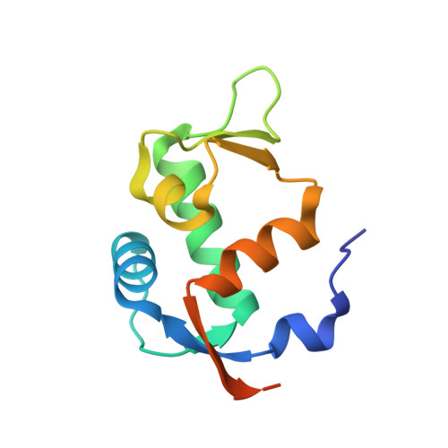
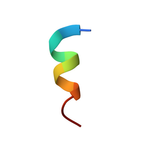
3JZR

Human MDM2 liganded with a 12mer peptide inhibitor (pDI6W)


Experimental Data Snapshot

  • Method: X-RAY DIFFRACTION
  • Resolution: 2.10 Å
  • R-Value Free: 0.228 
  • R-Value Work: 0.194 
  • R-Value Observed: 0.194 

Starting Model: experimental
View more details

wwPDB Validation   3D Report Full Report


This is version 1.2 of the entry. See complete history


Literature

Structure-based design of high affinity peptides inhibiting the interaction of p53 with MDM2 and MDMX.

Phan, J.Li, Z.Kasprzak, A.Li, B.Sebti, S.Guida, W.Schonbrunn, E.Chen, J.

(2010) J Biol Chem 285: 2174-2183

  • DOI: https://doi.org/10.1074/jbc.M109.073056
  • Primary Citation of Related Structures:  
    3JZO, 3JZP, 3JZQ, 3JZR, 3JZS

  • PubMed Abstract: 

    MDM2 and MDMX function as key regulators of p53 by binding to its N terminus, inhibiting its transcriptional activity, and promoting degradation. MDM2 and MDMX overexpression or hyperactivation directly contributes to the loss of p53 function during the development of nearly 50% of human cancers. Recent studies showed that disrupting p53-MDM2 and p53-MDMX interactions can lead to robust activation of p53 but also revealed a need to develop novel dual specific or MDMX-specific inhibitors. Using phage display we identified a 12-residue peptide (pDI) with inhibitory activity against MDM2 and MDMX. The co-crystal structures of the pDI and a single mutant derivative (pDI6W) liganded with the N-terminal domains of human MDMX and MDM2 served as the basis for the design of 11 distinct pDI-derivative peptides that were tested for inhibitory potential. The best derivative (termed pDIQ) contained four amino acid substitutions and exhibited a 5-fold increase in potency over the parent peptide against both MDM2 (IC(50) = 8 nm) and MDMX (IC(50) = 110 nm). Further structural studies revealed key molecular features enabling the high affinity binding of the pDIQ to these proteins. These include large conformational changes of the pDIQ to reach into a hydrophobic site unique to MDMX. The findings suggest new strategies toward the rational design of small molecule inhibitors efficiently targeting MDMX.


  • Organizational Affiliation

    Molecular Oncology, H. Lee Moffitt Cancer Center and Research Institute, Tampa, Florida 33612, USA.


Macromolecules
Find similar proteins by:  (by identity cutoff)  |  3D Structure
Entity ID: 1
MoleculeChains Sequence LengthOrganismDetailsImage
E3 ubiquitin-protein ligase Mdm2110Homo sapiensMutation(s): 0 
Gene Names: MDM2
EC: 6.3.2 (PDB Primary Data), 2.3.2.27 (UniProt)
UniProt & NIH Common Fund Data Resources
Find proteins for Q00987 (Homo sapiens)
Explore Q00987 
Go to UniProtKB:  Q00987
PHAROS:  Q00987
GTEx:  ENSG00000135679 
Entity Groups  
Sequence Clusters30% Identity50% Identity70% Identity90% Identity95% Identity100% Identity
UniProt GroupQ00987
Sequence Annotations
Expand
  • Reference Sequence

Find similar proteins by:  Sequence   |   3D Structure  

Entity ID: 2
MoleculeChains Sequence LengthOrganismDetailsImage
pDI6W peptide (12mer)B [auth P]12N/AMutation(s): 0 
Entity Groups  
Sequence Clusters30% Identity50% Identity70% Identity90% Identity95% Identity100% Identity
Sequence Annotations
Expand
  • Reference Sequence
Experimental Data & Validation

Experimental Data

  • Method: X-RAY DIFFRACTION
  • Resolution: 2.10 Å
  • R-Value Free: 0.228 
  • R-Value Work: 0.194 
  • R-Value Observed: 0.194 
  • Space Group: I 2 2 2
Unit Cell:
Length ( Å )Angle ( ˚ )
a = 37.223α = 90
b = 68.832β = 90
c = 92.391γ = 90
Software Package:
Software NamePurpose
StructureStudiodata collection
CNSrefinement
HKL-2000data reduction
HKL-2000data scaling
CNSphasing

Structure Validation

View Full Validation Report


Entry History 

Deposition Data

Revision History  (Full details and data files)

  • Version 1.0: 2009-11-10
    Type: Initial release
  • Version 1.1: 2011-07-13
    Changes: Version format compliance
  • Version 1.2: 2023-09-06
    Changes: Data collection, Database references, Refinement description