3OQF | pdb_00003oqf

Crystal Structure Analysis of Renin-indole-piperazine inhibitor complexes


Experimental Data Snapshot

  • Method: X-RAY DIFFRACTION
  • Resolution: 2.78 Å
  • R-Value Free: 
    0.201 (Depositor), 0.203 (DCC) 
  • R-Value Work: 
    0.180 (Depositor), 0.180 (DCC) 
  • R-Value Observed: 
    0.181 (Depositor) 

wwPDB Validation 3D Report Full Report

Validation slider image for 3OQF

Ligand Structure Quality Assessment 


This is version 1.3 of the entry. See complete history

Literature

Discovery and optimization of a new class of potent and non-chiral indole-3-carboxamide-based renin inhibitors.

Scheiper, B.Matter, H.Steinhagen, H.Stilz, U.Bocskei, Z.Fleury, V.McCort, G.

(2010) Bioorg Med Chem Lett 20: 6268-6272

  • DOI: https://doi.org/10.1016/j.bmcl.2010.08.092
  • Primary Citation Related Structures: 
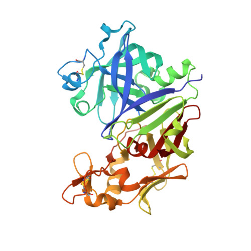
    3OOT, 3OQF, 3OQK

  • PubMed Abstract: 

    Selective inhibition of the aspartyl protease renin has gained attraction as an interesting approach to control hypertension and associated cardiovascular risk factors given its unique position in the renin-angiotensin system. Using a combination of high-throughput screening, parallel synthesis, X-ray crystallography and structure-based design, we identified and optimized a novel series of potent and non-chiral indole-3-carboxamides with remarkable potency for renin. The most potent compound 5k displays an IC(50) value of 2nM.


  • Organizational Affiliation
    • Sanofi-Aventis Deutschland GmbH, Chemical and Analytical Sciences, Building G878, D-65926 Frankfurt, Germany. Bodo.Scheiper@sanofi-aventis.com

Macromolecule Content 

  • Total Structure Weight: 75.77 kDa 
  • Atom Count: 5,479 
  • Modeled Residue Count: 674 
  • Deposited Residue Count: 680 
  • Unique protein chains: 1

Macromolecules

Find similar proteins by:|  3D Structure
Entity ID: 1
MoleculeChains  Sequence LengthOrganismDetailsImage
Renin
A, B
340Homo sapiensMutation(s): 0 
Gene Names: REN
EC: 3.4.23.15
UniProt & NIH Common Fund Data Resources
Find proteins for P00797 (Homo sapiens)
Explore P00797 
Go to UniProtKB:  P00797
PHAROS:  P00797
GTEx:  ENSG00000143839 
Entity Groups
Sequence Clusters30% Identity50% Identity70% Identity90% Identity95% Identity100% Identity
UniProt GroupP00797
Glycosylation
Glycosylation Sites: 1Go to GlyGen: P00797-1
Sequence Annotations
Expand
Reference Sequence

Small Molecules

Experimental Data & Validation

Experimental Data

  • Method: X-RAY DIFFRACTION
  • Resolution: 2.78 Å
  • R-Value Free:  0.201 (Depositor), 0.203 (DCC) 
  • R-Value Work:  0.180 (Depositor), 0.180 (DCC) 
  • R-Value Observed: 0.181 (Depositor) 
Space Group: P 21 3
Unit Cell:
Length ( Å )Angle ( ˚ )
a = 143.22α = 90
b = 143.22β = 90
c = 143.22γ = 90
Software Package:
Software NamePurpose
SCALEPACKdata scaling
BUSTER-TNTrefinement
PDB_EXTRACTdata extraction
CrystalCleardata collection
MOSFLMdata reduction
BUSTERrefinement

Structure Validation

View Full Validation Report


Ligand Structure Quality Assessment 


Entry History 

Deposition Data

  • Released Date: 2010-10-13 
  • Deposition Author(s): Bocskei, Z.

Revision History  (Full details and data files)

  • Version 1.0: 2010-10-13
    Type: Initial release
  • Version 1.1: 2011-07-13
    Changes: Version format compliance
  • Version 1.2: 2020-07-29
    Type: Remediation
    Reason: Carbohydrate remediation
    Changes: Data collection, Derived calculations, Structure summary
  • Version 1.3: 2024-10-09
    Changes: Data collection, Database references, Structure summary