3Q2X | pdb_00003q2x

Structure of an amyloid forming peptide NKGAII (residues 27-32) from amyloid beta


Experimental Data Snapshot

  • Method: X-RAY DIFFRACTION
  • Resolution: 1.45 Å
  • R-Value Free: 
    0.259 (Depositor), 0.265 (DCC) 
  • R-Value Work: 
    0.204 (Depositor), 0.207 (DCC) 
  • R-Value Observed: 
    0.209 (Depositor) 

wwPDB Validation 3D Report Full Report

Validation slider image for 3Q2X

This is version 1.3 of the entry. See complete history

Literature

Molecular basis for amyloid-beta polymorphism.

Colletier, J.P.Laganowsky, A.Landau, M.Zhao, M.Soriaga, A.B.Goldschmidt, L.Flot, D.Cascio, D.Sawaya, M.R.Eisenberg, D.

(2011) Proc Natl Acad Sci U S A 108: 16938-16943

  • DOI: https://doi.org/10.1073/pnas.1112600108
  • Primary Citation Related Structures: 
    2Y29, 2Y2A, 2Y3J, 2Y3K, 2Y3L, 3OW9, 3PZZ, 3Q2X

  • PubMed Abstract: 

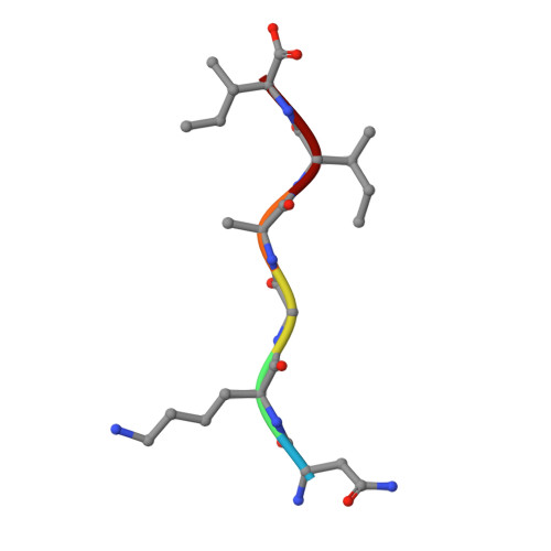
    Amyloid-beta (Aβ) aggregates are the main constituent of senile plaques, the histological hallmark of Alzheimer's disease. Aβ molecules form β-sheet containing structures that assemble into a variety of polymorphic oligomers, protofibers, and fibers that exhibit a range of lifetimes and cellular toxicities. This polymorphic nature of Aβ has frustrated its biophysical characterization, its structural determination, and our understanding of its pathological mechanism. To elucidate Aβ polymorphism in atomic detail, we determined eight new microcrystal structures of fiber-forming segments of Aβ. These structures, all of short, self-complementing pairs of β-sheets termed steric zippers, reveal a variety of modes of self-association of Aβ. Combining these atomic structures with previous NMR studies allows us to propose several fiber models, offering molecular models for some of the repertoire of polydisperse structures accessible to Aβ. These structures and molecular models contribute fundamental information for understanding Aβ polymorphic nature and pathogenesis.


  • Organizational Affiliation
    • Howard Hughes Medical Institute, Department of Biological Chemistry and Chemistry and Biochemistry, UCLA-DOE Institute for Genomics and Proteomics, University of California, Los Angeles, CA 90095, USA.

Macromolecule Content 

  • Total Structure Weight: 0.62 kDa 
  • Atom Count: 45 
  • Modeled Residue Count: 6 
  • Deposited Residue Count: 6 
  • Unique protein chains: 1

Macromolecules

Find similar proteins by:  Sequence   |   3D Structure  
Entity ID: 1
MoleculeChains  Sequence LengthOrganismDetailsImage
Amyloid beta A4 protein6Homo sapiensMutation(s): 0 
UniProt & NIH Common Fund Data Resources
Find proteins for P05067 (Homo sapiens)
Explore P05067 
Go to UniProtKB:  P05067
PHAROS:  P05067
GTEx:  ENSG00000142192 
Entity Groups
UniProt GroupP05067
Sequence Annotations
Expand
Reference Sequence

Experimental Data & Validation

Experimental Data

  • Method: X-RAY DIFFRACTION
  • Resolution: 1.45 Å
  • R-Value Free:  0.259 (Depositor), 0.265 (DCC) 
  • R-Value Work:  0.204 (Depositor), 0.207 (DCC) 
  • R-Value Observed: 0.209 (Depositor) 
Space Group: P 1 21 1
Unit Cell:
Length ( Å )Angle ( ˚ )
a = 15.074α = 90
b = 4.838β = 95.56
c = 24.016γ = 90
Software Package:
Software NamePurpose
DENZOdata reduction
SCALEPACKdata scaling
PHASERphasing
PHENIXrefinement
PDB_EXTRACTdata extraction

Structure Validation

View Full Validation Report


Entry History 

Deposition Data

Revision History  (Full details and data files)

  • Version 1.0: 2011-10-12
    Type: Initial release
  • Version 1.1: 2011-11-30
    Changes: Database references
  • Version 1.2: 2017-09-13
    Changes: Database references, Source and taxonomy, Structure summary
  • Version 1.3: 2024-02-21
    Changes: Data collection, Database references