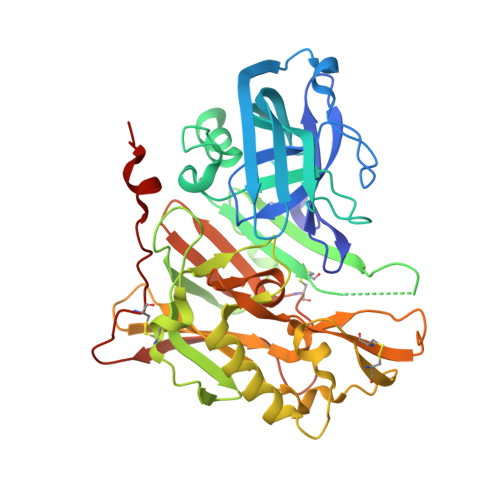
3UDH

Crystal Structure of BACE with Compound 1


Experimental Data Snapshot

  • Method: X-RAY DIFFRACTION
  • Resolution: 1.70 Å
  • R-Value Free: 0.241 
  • R-Value Work: 0.196 
  • R-Value Observed: 0.198 

wwPDB Validation   3D Report Full Report


Ligand Structure Quality Assessment 


This is version 1.2 of the entry. See complete history


Literature

Discovery and optimization of a novel spiropyrrolidine inhibitor of {beta}-secretase (BACE1) through fragment-based drug design.

Efremov, I.V.Vajdos, F.F.Borzilleri, K.A.Capetta, S.Chen, H.Dorff, P.H.Dutra, J.K.Goldstein, S.W.Mansour, M.McColl, A.Noell, S.Oborski, C.E.O'Connell, T.N.O'Sullivan, T.J.Pandit, J.Wang, H.Wei, B.Withka, J.M.

(2012) J Med Chem 55: 9069-9088

  • DOI: https://doi.org/10.1021/jm201715d
  • Primary Citation of Related Structures:  
    3UDH, 3UDJ, 3UDK, 3UDM, 3UDN, 3UDP, 3UDQ, 3UDR, 3UDY

  • PubMed Abstract: 

    The aspartyl protease β-secretase, or BACE, has been demonstrated to be a key factor in the proteolytic formation of Aβ-peptide, a major component of plaques in the brains of Alzheimer's disease (AD) patients, and inhibition of this enzyme has emerged as a major strategy for pharmacologic intervention in AD. An X-ray-based fragment screen of Pfizer's proprietary fragment collection has resulted in the identification of a novel BACE binder featuring spiropyrrolidine framework. Although exhibiting only weak inhibitory activity against the BACE enzyme, the small compound was verified by biophysical and NMR-based methods as a bona fide BACE inhibitor. Subsequent optimization of the lead compound, relying heavily on structure-based drug design and computational prediction of physiochemical properties, resulted in a nearly 1000-fold improvement in potency while maintaining ligand efficiency and properties predictive of good permeability and low P-gp liability.


  • Organizational Affiliation

    Pfizer Worldwide Research, Groton Laboratories, Eastern Point Road, Groton, Connecticut 06340, United States. ivan.efremov@pfizer.com


Macromolecules
Find similar proteins by:  (by identity cutoff)  |  3D Structure
Entity ID: 1
MoleculeChains Sequence LengthOrganismDetailsImage
Beta-secretase 1404Homo sapiensMutation(s): 0 
Gene Names: BACE1BACEKIAA1149
EC: 3.4.23.46
UniProt & NIH Common Fund Data Resources
Find proteins for P56817 (Homo sapiens)
Explore P56817 
Go to UniProtKB:  P56817
PHAROS:  P56817
GTEx:  ENSG00000186318 
Entity Groups  
Sequence Clusters30% Identity50% Identity70% Identity90% Identity95% Identity100% Identity
UniProt GroupP56817
Sequence Annotations
Expand
  • Reference Sequence
Small Molecules
Ligands 3 Unique
IDChains Name / Formula / InChI Key2D Diagram3D Interactions
091
Query on 091

Download Ideal Coordinates CCD File 
B [auth A](3S)-spiro[indole-3,3'-pyrrolidin]-2(1H)-one
C11 H12 N2 O
FQSHLSLQUHZOMV-LLVKDONJSA-N
ZN
Query on ZN

Download Ideal Coordinates CCD File 
E [auth A]ZINC ION
Zn
PTFCDOFLOPIGGS-UHFFFAOYSA-N
EDO
Query on EDO

Download Ideal Coordinates CCD File 
C [auth A]
D [auth A]
F [auth A]
G [auth A]
H [auth A]
C [auth A],
D [auth A],
F [auth A],
G [auth A],
H [auth A],
I [auth A]
1,2-ETHANEDIOL
C2 H6 O2
LYCAIKOWRPUZTN-UHFFFAOYSA-N
Experimental Data & Validation

Experimental Data

  • Method: X-RAY DIFFRACTION
  • Resolution: 1.70 Å
  • R-Value Free: 0.241 
  • R-Value Work: 0.196 
  • R-Value Observed: 0.198 
  • Space Group: C 2 2 21
Unit Cell:
Length ( Å )Angle ( ˚ )
a = 74.803α = 90
b = 103.75β = 90
c = 99.593γ = 90
Software Package:
Software NamePurpose
d*TREKdata scaling
d*TREKdata reduction
REFMACrefinement
PDB_EXTRACTdata extraction
MAR345data collection
autoBUSTERphasing

Structure Validation

View Full Validation Report


Ligand Structure Quality Assessment 


Entry History 

Revision History  (Full details and data files)

  • Version 1.0: 2012-04-18
    Type: Initial release
  • Version 1.1: 2013-06-19
    Changes: Database references
  • Version 1.2: 2017-11-08
    Changes: Refinement description