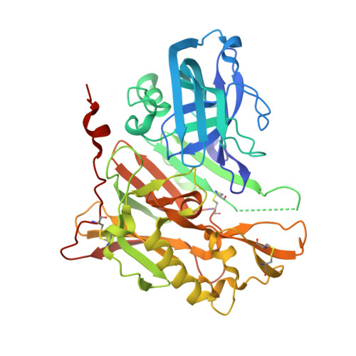
RCSB PDB - 3UDJ: Crystal Structure of BACE with Compound 5

 3UDJ

Crystal Structure of BACE with Compound 5


Experimental Data Snapshot

  • Method: X-RAY DIFFRACTION
  • Resolution: 1.80 Å
  • R-Value Free: 0.249 
  • R-Value Work: 0.207 
  • R-Value Observed: 0.209 

wwPDB Validation   3D Report Full Report


Ligand Structure Quality Assessment 

Created with Raphaël 2.3.0Worse 01 BetterLigand structure goodness of fit to experimental dataBest fitted 092Click on this verticalbar to view details

This is version 1.2 of the entry. See complete history


Literature

Discovery and optimization of a novel spiropyrrolidine inhibitor of {beta}-secretase (BACE1) through fragment-based drug design.

Efremov, I.V.Vajdos, F.F.Borzilleri, K.A.Capetta, S.Chen, H.Dorff, P.H.Dutra, J.K.Goldstein, S.W.Mansour, M.McColl, A.Noell, S.Oborski, C.E.O'Connell, T.N.O'Sullivan, T.J.Pandit, J.Wang, H.Wei, B.Withka, J.M.

(2012) J Med Chem 55: 9069-9088

  • DOI: https://doi.org/10.1021/jm201715d
  • Primary Citation of Related Structures:  
    3UDH, 3UDJ, 3UDK, 3UDM, 3UDN, 3UDP, 3UDQ, 3UDR, 3UDY

  • PubMed Abstract: 

    The aspartyl protease β-secretase, or BACE, has been demonstrated to be a key factor in the proteolytic formation of Aβ-peptide, a major component of plaques in the brains of Alzheimer's disease (AD) patients, and inhibition of this enzyme has emerged as a major strategy for pharmacologic intervention in AD. An X-ray-based fragment screen of Pfizer's proprietary fragment collection has resulted in the identification of a novel BACE binder featuring spiropyrrolidine framework. Although exhibiting only weak inhibitory activity against the BACE enzyme, the small compound was verified by biophysical and NMR-based methods as a bona fide BACE inhibitor. Subsequent optimization of the lead compound, relying heavily on structure-based drug design and computational prediction of physiochemical properties, resulted in a nearly 1000-fold improvement in potency while maintaining ligand efficiency and properties predictive of good permeability and low P-gp liability.


  • Organizational Affiliation

    Pfizer Worldwide Research, Groton Laboratories, Eastern Point Road, Groton, Connecticut 06340, United States. ivan.efremov@pfizer.com


Macromolecules
Find similar proteins by:  (by identity cutoff)  |  3D Structure
Entity ID: 1
MoleculeChains Sequence LengthOrganismDetailsImage
Beta-secretase 1404Homo sapiensMutation(s): 0 
Gene Names: BACE1BACEKIAA1149
EC: 3.4.23.46
UniProt & NIH Common Fund Data Resources
Find proteins for P56817 (Homo sapiens)
Explore P56817 
Go to UniProtKB:  P56817
PHAROS:  P56817
GTEx:  ENSG00000186318 
Entity Groups  
Sequence Clusters30% Identity50% Identity70% Identity90% Identity95% Identity100% Identity
UniProt GroupP56817
Sequence Annotations
Expand
  • Reference Sequence
Small Molecules
Ligands 4 Unique
IDChains Name / Formula / InChI Key2D Diagram3D Interactions
092
Query on 092

Download Ideal Coordinates CCD File 
B [auth A]methyl (3S,5'R)-2-oxo-1,2-dihydrospiro[indole-3,3'-pyrrolidine]-5'-carboxylate
C13 H14 N2 O3
WBRZAMFFUKWSQR-ZWNOBZJWSA-N
PEG
Query on PEG

Download Ideal Coordinates CCD File 
E [auth A]DI(HYDROXYETHYL)ETHER
C4 H10 O3
MTHSVFCYNBDYFN-UHFFFAOYSA-N
ZN
Query on ZN

Download Ideal Coordinates CCD File 
F [auth A]ZINC ION
Zn
PTFCDOFLOPIGGS-UHFFFAOYSA-N
EDO
Query on EDO

Download Ideal Coordinates CCD File 
C [auth A],
D [auth A]
1,2-ETHANEDIOL
C2 H6 O2
LYCAIKOWRPUZTN-UHFFFAOYSA-N
Experimental Data & Validation

Experimental Data

  • Method: X-RAY DIFFRACTION
  • Resolution: 1.80 Å
  • R-Value Free: 0.249 
  • R-Value Work: 0.207 
  • R-Value Observed: 0.209 
  • Space Group: C 2 2 21
Unit Cell:
Length ( Å )Angle ( ˚ )
a = 75.113α = 90
b = 104.016β = 90
c = 100.847γ = 90
Software Package:
Software NamePurpose
d*TREKdata scaling
d*TREKdata reduction
REFMACrefinement
PDB_EXTRACTdata extraction

Structure Validation

View Full Validation Report


Ligand Structure Quality Assessment 

Created with Raphaël 2.3.0Worse 01 BetterLigand structure goodness of fit to experimental dataBest fitted 092Click on this verticalbar to view details

Entry History 

Revision History  (Full details and data files)

  • Version 1.0: 2012-04-18
    Type: Initial release
  • Version 1.1: 2013-06-19
    Changes: Database references
  • Version 1.2: 2024-10-30
    Changes: Data collection, Database references, Derived calculations, Structure summary